
To combat the risks of radiolysis during high-activity production runs, the final formulation of the tracer now includes a 10 milligram per milliliter concentration of ascorbic acid. This critical adjustment ensures that the molecular integrity of the tracer remains intact even when using higher levels of gallium-68, which is essential for scaling up production to serve multiple patients from a single run. As of 2026, the landscape of oncology diagnostics is increasingly focused on the tumor microenvironment, specifically the role of cancer-associated fibroblasts that express fibroblast activation protein. This protein is highly prevalent in malignancies such as breast, ovarian, and colorectal cancers, yet it remains notably absent or minimal in healthy adult tissues, making it an exceptional target for high-contrast PET imaging. Single-domain antibodies, often referred to as nanobodies, have emerged as the preferred delivery vehicle for these applications due to their compact size of approximately 15 kilodaltons. Their small molecular footprint allows for exceptionally rapid blood clearance and deep penetration into tumor tissues, providing clinicians with high target-to-background ratios in a significantly shorter timeframe than traditional full-sized antibodies. The recent study published in EJNMMI Radiopharmacy and Chemistry marks a significant milestone by detailing how a previously preclinical protocol has been successfully transitioned into a robust, clinical-grade workflow suitable for modern hospital environments and centralized radiopharmacies.
1. Standardized Manufacturing Steps for PET Tracers
The transition from a laboratory setting to a clinical environment necessitated a rigorous standardization of the radiolabeling process to ensure repeatability and safety. Researchers identified that the core reaction requires a precise balance of temperature and chemical concentrations to maintain the stability of the antibody construct while maximizing the incorporation of the gallium-68 radionuclide. By using an ammonium acetate or sodium acetate buffer, the team maintained the required pH levels for optimal chelation. The precursor amount was strategically increased to 3 milligrams, allowing for the creation of multi-patient batches without sacrificing the molar activity needed for clear imaging. Furthermore, the protein dose administered to patients was set at 1 milligram, a level that has been shown to optimize the biodistribution of the tracer by reducing unnecessary liver uptake. This carefully tuned environment ensures that the chemical reaction proceeds efficiently during the ten-minute incubation period at 50 degrees Celsius, resulting in a product that consistently meets the stringent purity requirements of modern nuclear medicine departments.
The manufacturing workflow follows a specific sequence of operations designed to minimize human error and maximize radioactive yield. To facilitate clinical implementation, the following steps were established as the standard operating procedure: 1. Prepare the reaction environment: Elute gallium-68 using hydrochloric acid and combine it with 3 milligrams of the precursor in an acetate buffer solution. 2. Conduct the labeling incubation: Heat the mixture at 50 degrees Celsius for 10 minutes to facilitate the complexation of the radionuclide with the antibody construct. 3. Incorporate stabilizing agents: Add an ascorbic acid solution in saline to the mixture to prevent radiolysis and maintain product integrity. 4. Perform final sterile filtration: Pass the solution through a sterile filter to ensure clinical safety, followed by a saline rinse to recover any residual activity trapped in the system. This standardized sequence has proven to be highly effective, yielding radiochemical purity levels that frequently exceed 97 percent. The addition of the saline rinse step was particularly vital for automated systems, where a significant portion of the valuable tracer could otherwise remain stuck in the tubing or the filter membrane, potentially lowering the overall efficiency of the production run.
2. Optimizing Automated and Manual Platforms
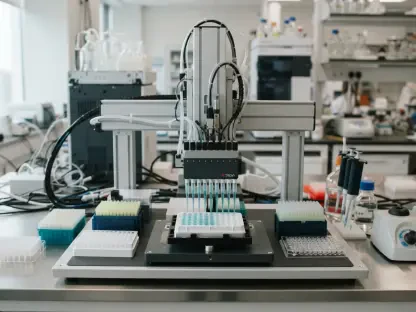
To ensure that this FAP-targeting tracer could be adopted by a wide variety of medical facilities, the researchers validated the production process on two prominent automated synthesis modules, the Modular-Lab Eazy and the Modular-Lab PharmTracer. These systems represent the current standard in automated radiochemistry, though they utilize different mechanical approaches for moving liquids. The Eazy platform relies primarily on controlled gas pressure to drive reagents through the disposable cassette, while the PharmTracer utilizes an automated syringe module for more precise volume control. Despite these mechanical differences, both platforms delivered remarkably consistent results after the introduction of a dedicated rinsing step. This modification was necessary because initial tests showed that a portion of the radiolabeled antibody was being lost during the final filtration process. By flushing the reaction vial and the sterile filter with 0.9 percent sodium chloride, the recovery rate was boosted to over 80 percent, while residual activity within the manifold was reduced to less than 2 percent. This level of efficiency is crucial for the economic viability of new tracers in a busy clinical setting.
Recognizing that not all imaging centers possess expensive automated synthesizers, the development team also perfected a manual, kit-based approach that offers similar performance without the need for complex hardware. This manual procedure involves the direct elution of gallium-68 into a reaction vial that already contains the necessary precursor and buffer components. The vial is then heated in a standard dry bath for ten minutes before being diluted with the ascorbic acid stabilizer and passed through a sterile filter. During validation runs, this manual method produced yields and purity levels that were virtually identical to those achieved by the automated platforms. This flexibility is further enhanced by the process’s compatibility with different generator systems, such as the GalliaPharma and Galli Eo models. Although these generators use different elution volumes of hydrochloric acid, the researchers demonstrated that simple adjustments to the buffer volume could maintain the ideal reaction conditions. This means that a facility’s choice of hardware does not limit its ability to produce high-quality FAP-targeting tracers, thereby democratizing access to this advanced imaging technology.
3. Ensuring Quality and Biological Function
A critical phase of the clinical translation involved a comprehensive stress study designed to identify the operational boundaries of the production process. By intentionally varying parameters such as reaction temperature, incubation time, and starting radioactivity, the research team established that the process is remarkably resilient but has specific limits. For instance, while the labeling efficiency remains high across a range of conditions, temperatures falling below 35 degrees Celsius result in incomplete chelation, leading to unacceptable levels of free gallium in the final product. Conversely, pushing the temperature above 60 degrees Celsius can be detrimental, as it may lead to protein precipitation or the formation of air bubbles that can block the sterile filters. The ten-minute incubation time was confirmed as the optimal window, providing enough time for the reaction to reach completion without allowing excessive radioactive decay to occur. The resulting product consistently presented as a clear, colorless solution with a pH between 4.5 and 5.0, meeting all physical and chemical criteria for human administration.
Beyond the chemical purity of the tracer, it was essential to verify that the manufacturing process did not damage the biological functionality of the single-domain antibody. The team employed advanced analytical methods, including enzyme-linked immunosorbent assays and bead-based binding tests, to confirm that the antibody’s affinity for the fibroblast activation protein remained intact. The results were impressive, with the relative binding potency exceeding 90 percent in all test cases, indicating that neither the heating nor the radiation exposure significantly altered the tracer’s ability to seek out tumor-associated fibroblasts. Stability studies further showed that the tracer remained within all quality specifications for at least three hours at room temperature, allowing for a comfortable window for quality control testing and transport to the patient. Endotoxin levels and microbiological assays consistently returned results below detection limits, confirming that the sterile filtration and aseptic handling procedures were fully effective. This rigorous validation provides clinicians with the confidence that the tracer will perform as expected in a diagnostic setting, providing clear and accurate images of the tumor microenvironment.
4. Operational Considerations for Molecular Imaging
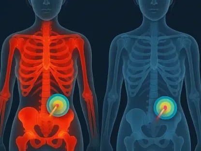
The implementation of this new process requires a clear understanding of the advantages offered by FAP-targeted imaging and the practical ways to maintain tracer quality. FAP is a highly significant target because of its role in the structural support of solid tumors; by imaging the fibroblasts rather than the cancer cells themselves, clinicians can gain a different perspective on the tumor’s extent and aggressiveness. Because the tracer uses a single-domain antibody, it avoids many of the pitfalls of larger proteins. These smaller molecules penetrate the dense extracellular matrix of the tumor more effectively, while their rapid clearance from the blood pool ensures that the “noise” in the image is minimized. This allows for high-contrast scans to be performed very soon after the tracer is injected, which is a major advantage for both patient comfort and clinic throughput. The compatibility of the process with various gallium-68 generators further simplifies the logistics, as hospitals can utilize their existing infrastructure to start producing the tracer without needing to purchase specific brand-name elution systems.
Maintaining the long-term viability of the tracer production also involves specific measures to prevent degradation and ensure safety during high-volume runs. The use of ascorbic acid at a concentration of 10 milligrams per milliliter is a proactive strategy to combat radiolysis, which is the breakdown of the tracer molecules caused by the energy of the radioactive decay itself. As centers look to increase the starting activity to produce enough for several patients simultaneously, the risk of radiolysis increases; the high concentration of ascorbic acid provides a robust chemical shield that preserves the tracer’s integrity throughout its shelf life. Additionally, the adherence to a strict ten-minute reaction time ensures that the ratio of labeled tracer to decayed isotope remains favorable. These operational details, when combined with the automated integrity tests performed by the synthesizers before each run, create a safety-first environment that is essential for routine clinical use. This comprehensive approach to manufacturing and quality control ensures that FAP-targeting PET imaging can move from a specialized research tool to a standard part of the oncological diagnostic toolkit.
5. Advancing Global Access to FAP Diagnostics
This research established a reliable framework for the clinical production of single-domain antibody-based radiopharmaceuticals, marking a definitive shift toward more accessible molecular imaging. The team determined that the transition from small-molecule tracers to protein-based agents did not have to result in overly complex or fragile manufacturing sequences. By mirroring the simplicity and robustness typically associated with simpler tracers, this process allowed for a seamless integration into existing hospital radiopharmacies. The consistent performance observed across different automated synthesizers and manual methods confirmed that the protocol was adaptable to the varied technical landscapes found in medical centers worldwide. This versatility ensured that the specific hardware available at a facility would not act as a barrier to providing patients with advanced FAP-targeted diagnostics, effectively broadening the reach of this technology beyond specialized research institutions.
The validation of these protocols provided a clear pathway for future developments in the field of nuclear medicine. The study demonstrated that high radiochemical yields and biological potency could be maintained even when scaling production for multi-patient use. This success encouraged the exploration of alternative chelators and room-temperature labeling techniques, which could further simplify the production of temperature-sensitive biological constructs. The researchers concluded that the integration of stabilizing agents and optimized rinsing steps was fundamental to achieving the high recovery rates necessary for clinical viability. As these processes become more commonplace, the framework established here served as a blueprint for the translation of other novel antibody-based tracers into the clinic. Ultimately, the work facilitated a more rapid adoption of FAP-targeted PET imaging, offering a practical solution for the high-volume production of sophisticated diagnostic agents in 2026 and beyond.