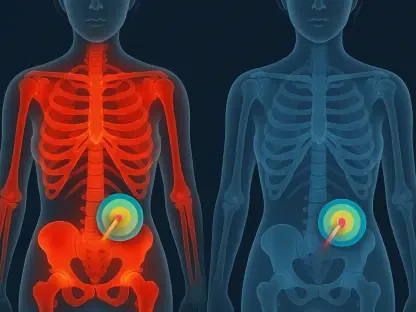

While T2-weighted sequences provide structural detail, the diffusion of water molecules within the tumor microenvironment remains a more sensitive indicator of vascular invasion. This physiological reality often presents a significant hurdle for oncologists trying to determine the best course of action for patients diagnosed with early-stage cervical cancer. Lymphovascular space invasion, or LVSI, represents a microscopic threshold where tumor cells begin to infiltrate the circulatory and lymphatic systems, marking a transition toward higher risk and more aggressive disease behavior. When LVSI is detected after surgery, it typically triggers a shift in the treatment paradigm, necessitating intensive postoperative interventions such as pelvic radiation therapy or chemotherapy to mitigate the risk of recurrence. Unfortunately, the current diagnostic standard often falls short, as conventional MRI lacks the spatial resolution necessary to see these cellular-level changes before the patient goes to the operating room. This creates a critical clinical gap where surgeons must operate without full knowledge of the tumor’s invasive potential, leading to either undertreatment or the need for secondary, stressful medical adjustments once pathology results return.
In an effort to resolve this ambiguity, researchers at the First People’s Hospital of Guangyuan have introduced a specialized diagnostic approach utilizing the power of deep learning to enhance traditional imaging. By applying a Super-Resolution Generative Adversarial Network, or SRGAN, to standard 3.0 Tesla MRI data, the medical team has successfully synthesized high-resolution representations that reveal the intricate textures of the tumor. This technological leap allows for a much more granular analysis of the tumor microenvironment, specifically focusing on tissue heterogeneity and the fine margins where invasion is most likely to occur. By bridging the gap between macroscopic imaging and microscopic pathology, this research offers a promising pathway for non-invasive, preoperative risk assessment. The ability to visualize these subtle markers of vascular invasion could revolutionize how gynecological cancers are staged, moving the field closer to a model where surgical and therapeutic plans are perfectly aligned with the biological reality of the patient’s condition before the first incision is ever made.
Clinical Context and Patient Demographics
The foundation of this investigation rested on a meticulously screened cohort of 117 female patients, all of whom were diagnosed with early-stage cervical cancer classified within the FIGO 2018 stages IB through IIB. These individuals were selected from a broader institutional database of over 1,300 records managed between 2026 and 2028, ensuring that the data reflected the most current clinical standards and patient management protocols. Every participant in the study underwent a total hysterectomy along with pelvic lymphadenectomy, a procedure that provided the essential histological gold standard required to verify the presence of lymphovascular space invasion (LVSI). By comparing the imaging data directly against these confirmed pathological results, the researchers established a high-fidelity baseline for their deep learning models. This rigorous selection process ensured that the resulting predictive framework was built on a solid empirical foundation, where every digital prediction could be cross-referenced with physical tissue samples analyzed by expert pathologists.
During the analysis of the final cohort, researchers observed distinct pathological differences between the 45 patients who tested positive for LVSI and the 72 who did not. While factors like age and primary tumor diameter remained relatively consistent across both groups, those with confirmed vascular invasion exhibited a significantly higher prevalence of pelvic lymph node metastasis and a much greater depth of cervical stromal invasion. These findings underscore the high-risk nature of LVSI-positive cases and highlight why early identification is vital for long-term survival. The presence of these aggressive features suggests that vascular invasion is not just an isolated finding but is often part of a broader, more dangerous progression of the disease. These data reinforce the urgent need for non-invasive predictive tools that can flag these high-risk indicators during the initial diagnostic workup, allowing clinicians to bypass the traditional wait for pathology results and move directly into a more targeted and comprehensive treatment strategy.
Imaging Protocols and Super-Resolution Reconstruction
The technological backbone of this research was a series of diagnostic scans conducted on a Siemens VIDA 3.0-Tesla scanner using specialized abdominal surface coils. The imaging protocol was designed to be comprehensive, incorporating axial T1-weighted and T2-weighted imaging with spectral attenuated inversion recovery, alongside diffusion-weighted imaging with varying b-values. This multi-sequence approach captured a diverse range of data, from structural anatomy to the minute movements of water molecules within the tissue. However, the true innovation occurred during the post-processing phase, where the researchers utilized a 3D SRGAN integrated into the OnekeyAI platform. Instead of relying solely on the original lower-resolution scans, the team used deep residual blocks to perform a four-fold spatial upsampling. This process refined the voxel dimensions of the diffusion-weighted imaging from 1.875 millimeters down to a crisp 0.469 millimeters, providing a level of detail that was previously inaccessible in routine clinical practice without significantly extending the patient’s time inside the imaging gantry.
What makes this super-resolution approach particularly effective is its use of a “frozen” inference engine, which leverages image priors from large-scale natural datasets to enhance medical scans. By applying these learned patterns to the specific anatomy of the cervix, the algorithm was able to sharpen the edges of the tumor and improve the overall granularity of the data. For the T2-weighted sequences, the resolution was enhanced from 0.781 millimeters to a mere 0.195 millimeters, creating an exceptionally clear view of the interface where the tumor meets the surrounding stroma. This is the exact region where microscopic vascular invasion typically begins, and having such a high-definition view allows for the extraction of much more descriptive radiomic features. This method demonstrates how AI can be used to “unlock” hidden information within standard scans, effectively upgrading the capabilities of existing hospital hardware through sophisticated software alone, thereby providing a more detailed diagnostic picture without increasing the financial or physical burden on the patient.
Objective Image Quality and Fidelity Assessments
To maintain scientific integrity and ensure the AI-generated images were accurate reflections of biological reality, the researchers conducted an exhaustive quality assessment. One of the primary concerns with any algorithmic reconstruction is the potential for “hallucinations,” where the system creates details that do not exist or introduces artifacts that could lead to an incorrect diagnosis. To counter this, the team measured the signal-to-noise ratio across a subset of patient scans, finding a measurable improvement in the super-resolution versions. For the diffusion-weighted images, the signal-to-noise ratio increased from 12.39 in the original files to 13.36 in the enhanced ones. More importantly, the standard deviation within the tumor regions remained stable throughout the process. This stability suggests that the algorithm successfully suppressed background noise and graininess without erasing the natural heterogeneity and internal texture of the tumor, which are the essential features needed for accurate radiomic modeling.
The T2-weighted sequences showed even more dramatic improvements, with the signal-to-noise ratio rising from 20.39 to over 22.00. While the researchers noted a slight smoothing effect, which is common in high-level algorithmic upsampling, the line-intensity profiles confirmed that the transitions across anatomical margins became significantly steeper. These sharper gradients are essential for clinicians because they define the exact boundary between cancerous tissue and healthy stroma with much greater precision. In the context of vascular invasion, where the difference between a contained tumor and an invasive one is measured in microns, the ability to see a clear, sharp margin is invaluable. These fidelity checks confirm that the super-resolution process does not just make images look “prettier” to the human eye but actually enhances the mathematical quality of the data, providing a more robust and reliable set of inputs for the subsequent machine learning stages of the diagnostic pipeline.
Radiomics Workflow and Feature Selection
The transition from raw images to predictive data began with a meticulous radiomics workflow, starting with the manual segmentation of the tumor volume. An experienced radiologist, working without knowledge of the final pathological results, used 3D Slicer software to delineate the regions of interest on a slice-by-slice basis. To ensure the highest level of data reliability, a subset of cases underwent a second round of segmentation after a month-long washout period, allowing for a rigorous intra-observer stability analysis. This check revealed that over 82 percent of the features derived from diffusion-weighted imaging were highly reproducible, achieving intraclass correlation coefficients of 0.75 or higher. In total, the team extracted 1,715 radiomic descriptors from the enhanced images, covering everything from basic shape and size to complex texture metrics that describe how pixels are distributed across the tumor’s three-dimensional volume.
Given the massive amount of data generated by this process, the next step involved sophisticated dimensionality reduction to isolate the most predictive signals. Using the training cohort alone to avoid any risk of information leakage, the researchers applied Spearman correlation filters to remove redundant features that were too closely related to one another. Following this, they utilized the Least Absolute Shrinkage and Selection Operator, better known as LASSO regression, to further prune the feature set. This statistical method is particularly effective at selecting a small, highly potent group of indicators while discarding the “noise” that can lead to model overfitting. By the end of this process, the researchers had distilled the thousands of original data points down to a handful of high-impact features. This refined dataset ensured that the final machine learning models were focused purely on the biological markers most closely associated with vascular invasion, resulting in a system that is both efficient and highly accurate.
Machine Learning Evaluation and Performance
With a refined set of features in hand, the researchers moved on to evaluate six different supervised machine learning architectures to see which could best predict the presence of LVSI. The lineup included well-known frameworks like Support Vector Machines, Logistic Regression, and K-Nearest Neighbors, alongside more advanced gradient boosting algorithms like XGBoost and LightGBM. Across nearly every test and validation metric, the XGBoost framework emerged as the clear leader. In the validation cohort, the model trained on super-resolution diffusion-weighted imaging achieved an area under the curve (AUC) of 0.864. This was a massive improvement over the model using original-resolution images, which only managed an AUC of 0.727. This statistically significant divergence proves that the added detail provided by the super-resolution process directly translates into a more powerful and reliable diagnostic tool for identifying complex pathological states.
While the structural T2-weighted models also showed gains after enhancement, the improvement was most profound in the diffusion-weighted sequences. This consistent upward trajectory in performance across all six architectures suggests that the benefits of super-resolution are robust and not just a fluke of one specific algorithm. Even the simpler models, such as the multilayer perceptron, saw their predictive power jump significantly when given access to the higher-resolution data. This broad success indicates that the “information density” of the scans was genuinely increased, providing the machine learning models with a more nuanced view of the tumor’s internal architecture. By successfully distinguishing between positive and negative LVSI cases with such high precision, these models demonstrate a level of diagnostic capability that far exceeds what is possible through traditional visual inspection of standard MRI scans, marking a significant step forward in oncological imaging.
Multimodal Fusion and Clinical Comparison
The culmination of the research was the development of a multi-sequence fusion model, which combined the most predictive features from both diffusion-weighted and T2-weighted scans. This integrated approach reached an impressive AUC of 0.934 in the independent validation cohort, demonstrating near-perfect diagnostic performance. To put this in perspective, the researchers compared the fusion model against a traditional model based only on clinical parameters, such as the patient’s age and the measured tumor diameter. The clinical-only model performed poorly by comparison, yielding an AUC of just 0.626. This stark contrast highlights that the most important markers of a tumor’s invasive potential are often hidden within its internal texture and microscopic behavior, rather than in external factors that are easily measured during a physical exam. Radiomic data essentially provides a “digital signature” of the tumor that is far more revealing than traditional clinical indicators.
One of the most surprising findings during the model’s development was that when clinical variables were added to the radiomic data, the statistical selection process actually excluded them as being redundant. This suggests that the high-dimensional features captured by the super-resolution imaging are so descriptive of the tumor’s biology that traditional markers, such as age, no longer add any useful information to the prediction. Decision curve analysis further validated the clinical utility of the SR-fusion model, showing that it provides the highest net benefit across a wide range of decision thresholds. For a surgeon deciding on the extent of a pelvic lymphadenectomy or a physician planning postoperative radiation, this model offers a level of confidence that was previously unattainable. By providing a clear, evidence-based prediction of vascular invasion, the technology allows for a more personalized and effective treatment plan, ensuring that high-risk patients receive the aggressive care they need while sparing lower-risk patients from unnecessary and potentially harmful overtreatment.
The Strategic Path Toward Implementing Virtual Biopsy Technologies and Advancing Personalized Patient Care Systems
The findings of this investigation confirmed that a 3D SRGAN-enhanced radiomics framework provides a superior method for the preoperative prediction of vascular invasion in cervical cancer. The core results showed that super-resolution processing not only increased the clarity of the images but also stabilized the radiomic features, making them more reliable across different models. The final SR-fusion model proved to be significantly more accurate than standard imaging or clinical assessments, offering a practical tool for risk stratification. It was observed that diffusion-weighted imaging provided the most stable and predictive data, suggesting that the movement of water molecules within the tumor is a key indicator of its invasive behavior. These developments established a new benchmark for how artificial intelligence can be integrated into the oncology workflow, providing a non-invasive way to see what was once only visible under a microscope.
Moving forward, this technology sets the stage for “virtual biopsy” techniques that could eventually reduce the reliance on invasive surgical procedures for staging. To build on this success, future research should focus on prospective, multicenter trials to ensure these models perform consistently across different types of MRI hardware and more diverse patient populations. There is also a need to transition from binary “yes-or-no” predictions to a more nuanced spectrum of risk, reflecting the clinical reality that vascular invasion can vary in intensity. By refining these AI tools, clinicians can gain a deeper understanding of each patient’s unique tumor biology, leading to highly tailored interventions that improve survival rates and quality of life. This research serves as a vital proof of concept, demonstrating that the future of cancer care lies in the intersection of advanced imaging, high-performance computing, and personalized medicine.