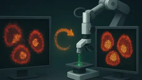

While Stochastic Optical Reconstruction Microscopy achieves nanoscopic precision, the quality of these images remains vulnerable to physical artifacts caused by inconsistent manual washing cycles. This fundamental discrepancy between advanced imaging hardware and manual preparation methods creates a significant bottleneck in modern biological research. Traditional protocols for techniques like STORM demand an exhaustive series of manual steps, ranging from precise cell fixation to the delicate application of fluorescent antibodies. Because these tasks rely heavily on the researcher’s steady hand, the potential for variability is immense. A single moment of aggressive pipetting or an uneven rinse can distort the very nanostructures a scientist aims to observe, leading to data that reflects human error rather than biological reality. By standardizing the fluidic environment, researchers can finally align the preparatory phase with the nanometric accuracy of the microscope itself, ensuring that every captured photon tells an authentic story of the cellular architecture.
Innovative Design and Performance Metrics
Engineering a Compact and Versatile Framework
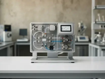
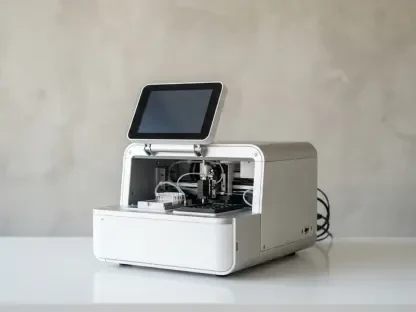
The Multi-Channel Automated Programmable Liquid Exchange System, or MAPLES, introduces a paradigm shift in laboratory efficiency through its remarkably compact and portable engineering. Measuring only 19 by 17 by 7 centimeters, the device occupies a footprint similar to a standard hardcover book, making it an ideal addition to even the most crowded laboratory benches. Weighing just 1.6 kilograms, the system is designed for mobility and rapid integration into existing microscopy setups without requiring permanent or intrusive modifications to high-end imaging platforms. This design philosophy directly addresses the limitations of previous automated solutions, which often consisted of either prohibitively expensive commercial units or bulky, home-built rigs that lacked the necessary refinement for delicate nanoscopic work. By prioritizing a small form factor, the engineers have ensured that high-precision automation is no longer a luxury reserved for massive core facilities but a tool accessible to research groups.
At the core of the MAPLES device is a sophisticated fluidic architecture that emphasizes cleanliness and the prevention of cross-contamination during complex staining procedures. Unlike traditional systems that route multiple reagents through a single shared line, this automated platform utilizes a dedicated syringe system for each specific liquid. This “disposable-first” approach is critical when working with sensitive biological reagents and expensive fluorescent markers that can be easily degraded or contaminated. By using air pressure to dispense fluids rather than direct mechanical contact with common pathways, the system maintains a sterile environment throughout the duration of a multiday experiment. Furthermore, the inclusion of a deliberate air gap above the sample bath ensures that reagents remain isolated, effectively eliminating the risk of carry-over between sequential labeling steps. This meticulous attention to fluid isolation guarantees that the chemical environment remains pristine for clarity.
Quantifying Volumetric Accuracy and Waste Reduction
Achieving the high standards of super-resolution microscopy requires an extreme degree of volumetric consistency that manual pipetting simply cannot provide over hundreds of repetitions. Quantitative analysis of the MAPLES platform demonstrates a remarkable liquid delivery error of only 3 percent during standard dispensing volumes. This level of precision is not just a momentary achievement but remains consistently stable across at least ten consecutive washing cycles, which is a common requirement for complex multiplexed staining protocols. By maintaining such tight tolerances, the automated system ensures that the concentration of buffers and antibodies remains uniform across every sample in a study, effectively removing a major source of experimental noise. This reliability allows researchers to compare data across different days or different laboratories with a newfound confidence that the results are a product of the biology being studied rather than fluctuations in reagent application or human performance.
Efficiency in reagent consumption is another critical metric where automation outperforms manual handling, particularly when dealing with specialized antibodies that are often prohibitively expensive. The MAPLES system features a minimized fluidic dead volume, typically ranging between 7 and 12 microliters, which ensures that precious reagents are not wasted in the internal plumbing of the device. Beyond conservation, the system also excels at waste removal, utilizing a high-precision aspiration mechanism that leaves less than 4 percent of residual fluid behind after each rinse. After just three successive automated cycles, the residual concentration of a previous reagent is reduced to a negligible 0.0064 percent, a level of purity that is nearly impossible to reach consistently with manual washing without risking sample damage. This rapid and thorough exchange of liquids is vital for preventing the background fluorescence that often obscures details. By optimizing delivery, the system maximizes the efficiency of every antibody microgram.
Biological Fidelity and Laboratory Standards
Preserving the Nanoscopic Architecture of Cells
To validate the biological fidelity of automated sample preparation, researchers conducted rigorous trials using human red blood cells, which are notoriously sensitive to environmental changes and mechanical stress. These cells lack internal support structures like a nucleus or traditional organelles, making their plasma membrane and underlying spectrin-actin cytoskeleton highly susceptible to damage. In experimental evaluations, the MAPLES system demonstrated its gentleness by keeping these fragile erythrocytes intact through twelve continuous automated rinse cycles at a high flow rate. Despite the volume of fluid passing over the cells, they remained firmly attached to the substrate without showing signs of rupture or morphological distortion. This success underscores the system’s ability to handle demanding biological specimens that would typically fail under the variable conditions of manual labor. By preserving the delicate cell boundary, automation confirms it can maintain the structural integrity.
The nanoscopic accuracy of the automated process was further confirmed through STORM imaging of the membrane-skeleton protein tropomodulin within the preserved red blood cells. The resulting high-resolution maps revealed an intricate distribution of protein clusters with a density of 70 units per square micrometer and a spacing of 80 nanometers. This data perfectly matched the known hexagonal lattice structure of the erythrocyte cytoskeleton, providing definitive proof that the automated cycles did not disrupt the molecular organization of the cell. In contrast, manual preparation often yields disorganized clusters that make it difficult to resolve underlying geometric patterns. The ability of the MAPLES system to consistently recover these nanometric details demonstrates that automation is not just a matter of convenience but a necessary technical requirement for high-fidelity mapping. These findings establish a new benchmark for reproducibility, ensuring that observations made at the nanoscale are accurate.
Establishing Reproducibility in Molecular Research
The development and implementation of the MAPLES system represented a transformative shift in the way super-resolution microscopy was approached within the laboratory. By replacing the variable nature of manual pipetting with a programmable, automated protocol, researchers successfully established a new standard for reproducibility in nanoscopic research. This transition ensured that the critical washing and staining phases—once considered the most vulnerable steps in a study—were executed with the same level of precision as the imaging itself. The system demonstrated that removing human variability from fluid handling directly improved the reliability of molecular data, allowing for more confident interpretations of cellular structures at the 20-nanometer scale. As laboratories adopted these automated tools, the focus of research shifted away from the mechanical challenges of sample preparation and toward the complex biological questions that super-resolution techniques were originally designed to answer.
Moving forward, the primary takeaway for the scientific community is the necessity of integrating automation into every stage of the imaging pipeline to ensure long-term data integrity. To fully capitalize on these technological gains, research institutions should prioritize the standardization of automated protocols, ensuring that digital scripts for fluid exchange are shared as openly as the imaging data itself. This move toward “programmable biology” will minimize the occurrence of preparation-induced artifacts and foster a culture of transparency and accuracy. Future research should also focus on integrating real-time sensor feedback into automated systems, allowing the hardware to adjust reagent delivery based on the immediate biochemical state of the sample. By treating sample preparation as a high-precision engineering task rather than a manual art form, scientists can ensure that the windows they open into the nanoscopic world remain clear and provide consistent results globally.