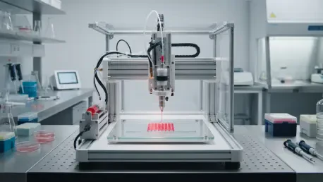

Recent advancements in CRISPR-Cas9 gene editing are being applied to create invisible cells that the human immune system will not target after transplantation. This critical innovation addresses one of the most persistent bottlenecks in regenerative medicine: the immune response that leads to the rejection of foreign tissues. The current landscape of organ transplantation is plagued by a widening disparity between the massive clinical demand for donor tissues and the limited biological supply available through traditional donation systems. While surgical transplantation remains the primary intervention for patients with end-stage organ failure, the reliance on human donors often results in long waiting lists and high mortality rates. This systemic crisis has catalyzed a decisive move toward the development of 3D bioprinting as a transformative methodology for creating functional organ substitutes. Unlike earlier tissue engineering approaches that were limited by static, prefabricated scaffolds, modern bioprinting enables the automated fabrication of complex, three-dimensional structures with anatomical precision. By depositing living cells and biomaterials layer by layer, this technology allows for the creation of intricate biological architectures that were previously impossible to replicate in a laboratory setting. This progress represents a fundamental shift toward personalized medicine, where replacement tissues are designed specifically for the unique anatomical and genetic needs of each individual patient.
Primary Modalities for Dispensing Biological Materials
The technical success of any bioprinting procedure depends heavily on the physical modality used to dispense biological materials into a structured format. Inkjet-based bioprinting has emerged as a high-precision, non-contact method that operates on a “drop-on-demand” principle. This system utilizes either thermal microheaters or piezoelectric crystals to generate pressure pulses that eject picoliter droplets of bioink through a fine nozzle. Because of its high spatial resolution, typically ranging between 20 and 100 micrometers, inkjet printing is exceptionally effective for creating delicate tissues that require exact cell placement. One of the primary advantages of this method is the high level of cell viability it maintains, often exceeding 85 percent, because the droplets are ejected with minimal mechanical force. However, the technology is generally restricted to low-viscosity bioinks, as thicker materials can lead to nozzle clogging and inconsistent droplet formation. Despite these limitations, the speed and accuracy of inkjet systems make them a vital tool for high-throughput applications where precision at the cellular level is the priority.
In contrast to droplet-based systems, extrusion-based bioprinting serves as the primary methodology for building robust, large-scale anatomical structures. This system utilizes pneumatic pressure or mechanical screws to dispense a continuous filament of material, allowing for the construction of dense, load-bearing tissues. Extrusion bioprinting is uniquely capable of handling high-viscosity hydrogels and shear-thinning materials, which are essential for maintaining the structural integrity of complex organs like the heart or liver. While the mechanical forces involved in extrusion can lower cell viability compared to inkjet methods, usually resulting in a range between 40 and 80 percent, the technique is indispensable for depositing high densities of cells. By allowing for the integration of multiple printheads, extrusion systems can simultaneously deposit different cell types and structural polymers, facilitating the creation of heterogeneous tissue constructs. This versatility has established extrusion as the workhorse of the industry, particularly for researchers focusing on orthopedic repair and the development of mechanically stable scaffolds for bone and cartilage.
Precision Through Light and Nozzleless Systems
To bypass the mechanical limitations associated with physical nozzles, researchers have increasingly adopted laser-assisted bioprinting and stereolithography. Laser-assisted bioprinting utilizes a pulsed laser focused onto a donor slide coated with a layer of bioink. The laser energy generates a vapor bubble that propels a precise droplet of the material onto a receiving substrate. This nozzle-free approach effectively eliminates the shear stresses that typically damage cells in extrusion systems, leading to exceptional cell viability levels that often surpass 90 percent. The resolution achieved through laser-assisted methods is incredibly fine, reaching as low as 10 micrometers, which allows for the replication of the most intricate biological patterns found in human tissues. Although the equipment and setup for laser-assisted systems are more complex and expensive than traditional printers, the ability to print cells without the risk of clogging or mechanical damage provides a significant advantage for the fabrication of delicate vascular networks and neural structures.
Stereolithography offers another high-resolution alternative by using light to selectively solidify liquid photopolymer resins. Guided by sophisticated computer-aided design models, a light source—either ultraviolet or visible light—crosslinks the bioink in a layer-by-layer fashion to build the desired structure. This process is prized for its ability to produce tissues with smooth surface finishes and complex internal geometries, making it ideal for replicating the curved surfaces of joints or the delicate structures of the eye. Because stereolithography does not rely on mechanical extrusion, it can produce highly detailed parts with a resolution and speed that mechanical systems struggle to match. However, the methodology requires the development of specialized photocurable bioinks that are both biocompatible and capable of rapid solidification. Careful management of light intensity is also necessary to prevent phototoxicity, which can harm the living cells embedded within the resin. Despite these challenges, stereolithography remains a leading choice for the precision engineering of cartilage and other tissues that require high structural definition.
The Evolution of Multi-Functional Bioink Formulations
The performance of any bioprinted construct is fundamentally tied to the properties of the bioink, which must serve as both a protective delivery vehicle and a supportive growth environment. Hydrogels are the most common foundation for bioinks due to their high water content, which effectively mimics the natural extracellular matrix of human tissues. Natural polymers like collagen, gelatin, and fibrin are highly valued in this context because they contain inherent biological signals that encourage cells to adhere, migrate, and proliferate. Alginate, a seaweed-derived polymer, is also frequently used because it can be rapidly crosslinked into a solid gel when exposed to calcium ions, providing immediate structural stability post-printing. However, while natural polymers offer excellent biological compatibility, they often lack the mechanical strength required for permanent implantation in weight-bearing areas of the body. This limitation has led to a growing focus on the optimization of polymer concentrations and the development of specialized additives that can enhance the physical properties of natural hydrogels without compromising their biological benefits.
To address the mechanical shortcomings of natural materials, the field has moved toward the use of hybrid and synthetic bioink formulations. Synthetic polymers like polycaprolactone and polyethylene glycol provide the structural backbone necessary for tissues that must withstand significant physical stress, such as bone or heart valves. These materials can be precisely engineered to have specific degradation rates, ensuring that the scaffold remains intact until the body’s own cells have replaced it with native tissue. Modern researchers are now creating composite inks that combine the biological advantages of natural substances with the mechanical robustness of synthetics, often incorporating nanoparticles to improve conductivity or structural reinforcement. This hybrid approach allows for the creation of bioinks that are shear-thinning during the printing process but become rigid and stable once they have been deposited. By tailoring the rheological and mechanical properties of these materials, bioengineers can now design tissues that are both biologically active and physically durable, moving the technology closer to widespread clinical application.
Navigating the Workflow From Design to Maturation
The creation of a bioprinted organ involves a sophisticated digital-to-biological workflow that begins with precise anatomical imaging. Using high-resolution scans from magnetic resonance imaging or computed tomography, engineers create a detailed 3D model of the patient’s defect or organ. This digital blueprint is then translated into a series of printing instructions, or “G-code,” which guides the motion of the bioprinter’s nozzles. Before printing begins, the bioink must be carefully tuned to ensure its flow characteristics match the intended resolution and speed of the print. This calibration phase is critical because even minor fluctuations in pressure or temperature can result in structural failure or cellular death. Once the printing process is initiated, the printer deposits the cellular material onto a sterile substrate, building the tissue layer by layer. The precision of this automated process allows for the strategic placement of different cell types in specific locations, mimicking the natural heterogeneity of human organs and ensuring that the final product is as functional as possible.
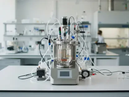
Once the physical structure has been printed, it enters the critical maturation phase, where it is placed inside a bioreactor to develop into living tissue. A bioreactor is a specialized device that provides a controlled environment mimicking the conditions of the human body, including optimal temperature, pH, and oxygen levels. Beyond just maintaining life, the bioreactor plays an active role in “training” the tissue through mechanical, electrical, or chemical stimulation. For instance, a printed heart valve may be subjected to pulsatile fluid flow to encourage the cells to align and strengthen the tissue, while a bone scaffold might receive mechanical compression to promote mineralization. This maturation process is essential because a freshly printed structure is merely a collection of cells and gel; it is the time spent in the bioreactor that allows the cells to form connections and produce their own extracellular matrix. Successful maturation ensures that the engineered tissue achieves the functional maturity required to survive and integrate into the patient’s body after the final transplantation procedure.
Solving the Critical Challenge of Tissue Vascularization
The primary obstacle preventing the fabrication of full-sized organs like the liver or heart is the “diffusion limit,” which restricts the thickness of engineered tissues. Without an integrated network of blood vessels, cells located more than a few hundred microns from the surface of a tissue construct cannot receive the oxygen and nutrients they need to survive, nor can they effectively remove waste products. This leads to localized necrosis in the center of the printed tissue, rendering large-scale constructs non-functional. To solve this, bioengineers are developing innovative techniques to print vascular channels directly into the tissue structure. One prominent approach involves the use of “sacrificial inks,” which are materials printed to occupy the space of future blood vessels. After the surrounding tissue has been stabilized through crosslinking, the sacrificial material is dissolved or melted away, leaving behind a network of hollow channels. These channels can then be lined with endothelial cells to create a perfusable circulatory system that can be connected to the patient’s blood supply.
Beyond sacrificial materials, the integration of multiple cell types during the initial printing phase has shown significant promise for the spontaneous formation of blood vessels. By co-printing endothelial cells with supporting mural cells and specialized growth factors, researchers are encouraging the biological process of angiogenesis within the engineered construct. This method relies on the innate ability of cells to organize themselves into tubes and networks when provided with the correct environment and signals. The combination of pre-printed larger vessels and biologically driven microvascular networks is currently seen as the most viable path toward creating thick, metabolically active organs. Furthermore, the use of advanced microfluidic bioreactors allows for the continuous perfusion of these nascent vascular systems during the maturation phase, ensuring that the interior of the organ remains viable as it grows. Solving the vascularization problem is the final technical hurdle that will enable 3D bioprinting to transition from producing thin tissue patches to manufacturing fully functional, life-saving organ replacements.
Future Perspectives on Implementation and Compliance
The advancement of 3D bioprinting necessitated a coordinated effort between bioengineers and regulatory agencies to ensure the safety of these complex therapies. Researchers integrated artificial intelligence systems that monitored the printing process in real-time, allowing for the automatic correction of errors and the optimization of cell distribution. Stakeholders established robust pipelines for clinical-grade cell sourcing and prioritized the automation of bioreactor systems to maintain consistency across large-scale manufacturing. These protocols focused on validating the mechanical integrity and biological compatibility of every printed construct, ensuring that patients received tissues that were free from contamination and optimized for long-term survival. The transition toward clinical implementation required the establishment of Good Manufacturing Practice standards specifically tailored for living, printed products. By aligning technological innovation with rigorous safety oversight, the medical community created a clear roadmap for the deployment of complex organ substitutes, transforming the landscape of surgical intervention and patient care.
The evolution of bioprinting also involved the successful integration of 4D bioprinting, where “smart” materials were used to create tissues that adapted to the patient’s body over time. This approach allowed for the fabrication of constructs that could change their shape or function in response to internal triggers, such as changes in pH or mechanical stress. Doctors utilized these dynamic materials to create growth-accommodating implants for pediatric patients, effectively reducing the need for multiple follow-up surgeries. Regulatory bodies like the FDA and EMA categorized these bioprinted tissues as high-risk combination products, leading to a decade of extensive clinical trials that confirmed their efficacy in treating chronic conditions. These efforts shifted the paradigm of medicine from managing organ failure to actively repairing and replacing damaged biological systems with precision-engineered solutions. The successful demonstration of vascularized skin and bone grafts provided the foundational data needed to support more ambitious projects, ultimately paving the way for the routine production of patient-specific organ replacements.