
The biological window for non-surgical jaw expansion is narrow, making the accurate timing of orthodontic intervention a critical factor for long-term patient outcomes. Maxillary expansion is a staple of pediatric dentistry, but the threshold between a simple mechanical procedure and a complex surgical requirement remains difficult to pinpoint. Recent advancements in deep learning, particularly the YOLOv11 architecture, have introduced a level of objectivity that was previously unattainable. This transition from manual radiographic inspection to automated feature extraction represents a fundamental shift in how practitioners evaluate skeletal maturity. By utilizing the specific patterns found in cone-beam computed tomography, artificial intelligence can now act as a high-fidelity filter, distinguishing between various stages of ossification that often appear identical to the human eye. This progress ensures that the treatment window is utilized effectively, reducing the risk of late-stage complications and improving the overall success rate of non-invasive procedures.
The DilemmSubjective Assessment in Modern Practice
Accurately staging the maturity of the midpalatal suture is vital because an incorrect guess has real-world consequences for the patient. If an orthodontist attempts to expand a fused suture, the patient may experience significant pain, and the teeth might tip outward rather than the jaw bone widening correctly. Conversely, referring a patient for invasive surgery when their suture is still open adds unnecessary cost, psychological stress, and recovery time to their treatment plan. The clinical environment demands a level of precision that manual review occasionally fails to provide, especially when dealing with the high volume of cases seen in modern clinics. This lack of certainty often forces practitioners to take a more conservative or aggressive approach than necessary, highlighting the limitations of human observation in detecting microscopic bony changes. The demand for a more refined diagnostic tool has pushed researchers to look toward computational solutions that can offer consistency across various patient demographics.
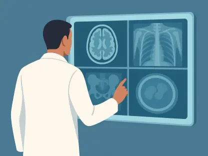
The current gold standard, known as the Angelieri classification system, categorizes suture maturation into five distinct stages. Despite its widespread use, the gray areas between these stages often lead to significant inter-observer variability, even among seasoned professionals. Practitioners frequently struggle to distinguish between partial fusion and total closure in complex cases where the bone density varies. This inconsistency highlights a desperate need for an objective, automated tool that can provide a reliable second opinion in a fast-paced clinical setting. Furthermore, the variability in scan quality from different imaging centers complicates the matter, as contrast and noise can obscure the subtle landmarks required for an accurate manual reading. By automating this classification, the industry can move toward a standardized protocol where the diagnosis is based on mathematical certainty rather than visual estimation. This shift is expected to reduce the incidence of failed mechanical expansions from 2026 to 2028 as these tools become more common.
The Technological Core: How YOLOv11x Transforms Detection
To address these diagnostic hurdles, researchers have turned to YOLOv11x, which represents the most robust version of the “You Only Look Once” deep learning framework. Unlike older AI models that analyze images in multiple steps—often losing anatomical context in the process—YOLOv11x processes an entire image in a single forward pass. This efficiency is critical in medical imaging because it allows the network to understand the global context of the anatomy, ensuring that subtle bony bridges are not viewed in isolation. The speed of this architecture means that a diagnosis can be rendered in milliseconds, allowing for a seamless integration into the diagnostic workflow. For the orthodontist, this means that the results of a scan can be discussed with the patient immediately during the initial consultation. This real-time capability is a significant improvement over previous generations of AI that required heavy cloud computing and lengthy processing times to reach a conclusion.
The “x” or “extra-large” variant of the model was specifically chosen for its superior feature extraction capabilities and high parameter count. By utilizing this high-capacity architecture, the study aimed to see if the AI could not only find the suture within a complex three-dimensional scan but also categorize it into clinically actionable groups with the precision of a master radiologist. This technological leap represents a shift from simple image recognition to sophisticated anatomical interpretation, where the software identifies the unique textures of ossified tissue. The model utilizes advanced attention mechanisms to focus on the narrow corridor of the midpalatal suture, effectively ignoring the surrounding dental noise that often distracts human observers. By isolating these specific biological markers, YOLOv11x achieves a level of granularity that was previously impossible. This deep learning approach allows the system to learn from thousands of variations in human anatomy, eventually outperforming the narrow experience of a single human practitioner.
Data Integrity: Building a Robust Foundation for Validation
The credibility of this AI model rests on a substantial dataset of 1,501 cone-beam computed tomography examinations from patients aged 10 to 25. This specific age range is crucial because it captures the most critical biological window for jaw growth and the subsequent fusion of the facial bones. To make the model’s findings more useful for doctors, the researchers condensed the five traditional Angelieri stages into three practical, actionable groups. These included early-stage open sutures, intermediate partial fusion, and late-stage fused sutures. By simplifying the output, the researchers ensured that the AI provided clear guidance on whether to proceed with expansion or prepare for surgery. The volume of data used in training allowed the neural network to identify rare anatomical variations that might only be seen a few times in a typical clinician’s career. This broad exposure ensures that the model remains accurate even when presented with unusual or asymmetric growth patterns in adolescent patients.
One of the most impressive aspects of the study was the focus on cross-device reliability, which is often a major hurdle for medical software. While the model was trained on images from one brand of scanner, it was tested on an entirely different hardware system to prove its versatility. This step is essential for proving that the technology can work in any dental office, regardless of the specific imaging equipment they own. It ensures the AI is identifying biological signatures rather than just memorizing the quirks of a specific camera or reconstruction algorithm. By testing on the J. Morita system after training on Planmeca hardware, the researchers demonstrated that the model could handle variations in noise, resolution, and contrast. This level of generalizability is what will allow the software to be deployed globally, supporting clinicians in various regions who use a wide array of technological tools. It bridges the gap between high-end research facilities and everyday clinical environments.
Performance Metrics: Quantifying the Impact of Deep Learning
When tested against unseen data from the second scanner, the YOLOv11x model achieved a mean average precision of 0.910. This high score indicates that the model is exceptionally talented at both locating the suture and correctly identifying its stage of growth. The system performed best at the two extremes—identifying clearly open sutures and clearly fused ones—which are the most critical data points for deciding on a treatment path. This high level of precision at the edges of the spectrum provides clinicians with the confidence they need to make definitive calls for or against surgical intervention. By reducing the number of false positives in the early stages, the AI ensures that patients who are eligible for non-invasive treatment do not undergo unnecessary surgery. Similarly, by accurately flagging fused sutures, it prevents the physical trauma associated with failed expansion attempts. This balance of sensitivity and specificity is what makes the model a viable clinical companion.
The performance in the intermediate Group C was slightly lower, mirroring the same difficulties faced by human doctors in everyday practice. This stage is characterized by ambiguous bony bridges that are difficult to see even on high-resolution scans, representing a biological transition zone. However, maintaining an overall accuracy of nearly 85 percent across different hardware brands proves that the AI is a robust tool that offers a level of consistency human observation cannot match over hundreds of cases. Even in these gray areas, the AI provided a probability score that could help clinicians weigh the risks of various treatment options. The ability of the model to maintain high performance across different manufacturers suggests that the underlying feature extraction is focusing on true skeletal maturation. As the software continues to evolve from 2026 to 2027, further refinement of these intermediate stages will likely be a primary focus for developers. This constant improvement cycle ensures that the tool remains at the cutting edge of orthodontic science.
The Path Forward: Clinical Integration and Evolution
While the results are promising, the transition from a research study to a daily clinical tool involves overcoming a few more practical hurdles. Currently, the model requires a human to select specific axial slices from a three-dimensional volume to ensure the AI is looking at the correct anatomical plane. The next step in this technological evolution would be for the AI to navigate the entire 3D scan automatically, picking the best angle for analysis without any human intervention. This would further reduce the time required for a diagnosis and eliminate the potential for human error during the slice selection process. Researchers are already looking into volumetric analysis where the AI calculates the total density of the suture across multiple planes. Such an approach would provide a more holistic view of the facial structure, accounting for the fact that fusion does not always happen uniformly from front to back. This leap into fully autonomous 3D analysis is the next logical step for the industry.
The researchers identified that the integration of automated diagnostics reduced the cognitive load on practitioners while increasing the reliability of patient screening protocols. This transition toward AI-driven analysis suggested that the next phase of orthodontic evolution would involve real-time volumetric scanning. Clinics that adopted these early iterations of the YOLOv11 framework observed a significant decrease in failed expansion attempts, validating the shift toward data-heavy decision-making. Future developments focused on expanding these neural networks to encompass broader demographics and varied ethnic anatomical markers. By moving away from subjective visual grading, the field moved into a more scientific era where treatment outcomes became more predictable and less dependent on individual clinician experience. This shift successfully bridged the gap between complex radiographic data and practical chairside application, ensuring that the technology remained a tool for empowerment rather than just a novelty in the digital landscape.