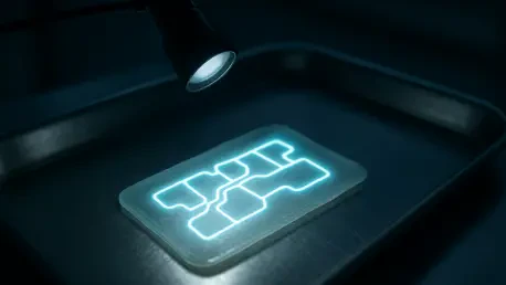

The design flexibility of bioprinted vascular platforms allows for the customization of cell ratios and geometries to meet the specific needs of individual patients. This high level of precision is particularly vital in 2026, as the medical community continues to face the mounting challenge of treating complex ischemic vascular diseases. These conditions, which occur when arterial blockages or narrowing restrict the supply of oxygen-rich blood to tissues, often result in debilitating outcomes such as critical limb ischemia. While conventional treatments including bypass surgery and stenting have provided relief for many, they are frequently insufficient for patients with extensive microvascular damage or those who lack suitable donor vessels for grafting. The emergence of 3D bioprinted human vascular organoid sheets, or hVOS, offers a sophisticated alternative that moves beyond mechanical plumbing to provide a biological solution capable of self-integration and active healing within a living host environment.
The Foundation: Engineering and Bioprinting
Advanced Bioprinting: Precision in Cellular Composition
The creation of human vascular organoid sheets begins with the strategic selection and preparation of specialized cell types derived from human pluripotent stem cells. Two primary components are essential for the architectural integrity of these tissues: endothelial cells and smooth muscle cells. Endothelial cells serve as the critical interior lining of the vascular system, facilitating the exchange of nutrients and signaling molecules, while smooth muscle cells provide the necessary structural support and contractile power to regulate blood pressure and flow volume. By using extrusion-based 3D bioprinting technology, researchers can deposit these cells at highly specific ratios within a biocompatible bioink. This bioink, often composed of gelatin methacryloyl, acts as a temporary scaffold that supports the cells during the printing process while maintaining a mechanical environment that encourages natural cellular activities.
This bioprinting methodology represents a significant advancement over earlier organoid cultivation techniques, which often suffered from unpredictable spatial arrangements and high variability between batches. In the current landscape of 2026, reproducibility is the cornerstone of clinical translation, and the ability to control the exact placement of thousands of cells ensures that each vascular sheet meets strict quality standards. The specialized bioink is engineered to be photocrosslinkable, allowing the structure to be stabilized instantly after printing. This structural stability provides the cells with the necessary time to establish their own internal biological connections without the risk of the scaffold collapsing. By standardizing these chemical and mechanical parameters, the engineering team has successfully mitigated the inconsistencies that once hindered the widespread application of stem-cell-derived vascular tissues.
Self-Assembly: The Emergence of Biological Complexity
Following the initial printing phase, the vascular sheets undergo a remarkable transition from a manufactured construct to a living, self-organizing tissue. Within the protective matrix of the gelatin-based scaffold, the endothelial and smooth muscle cells do not remain in their original printed locations; instead, they exhibit dynamic behaviors characterized by migration and spontaneous sorting. Over several days of in vitro culture, these cells communicate through biochemical signaling to form intricate, interconnected networks that mirror the complexity of genuine human vasculature. This process of self-assembly allows the tissue to develop mature features, such as tight junctions between endothelial cells and the formation of hollow, vessel-like lumens that are capable of carrying fluid.
To understand the internal mechanisms driving this maturation, the research team utilized high-resolution single-cell RNA sequencing to monitor the genetic activity of the cells over time. The data revealed that as the cells organized themselves into networks, they activated specific transcriptional programs related to mechanotransduction and hypoxic adaptation. Essentially, the cells were sensing the physical stresses of their environment and adjusting their genetic expression to prepare for the physiological demands they would face once transplanted into a living body. This biological “priming” is a critical step, as it ensures the cells are robust enough to survive the transition from a controlled laboratory incubator to the highly variable environment of an injured limb. This inherent ability of the hVOS to mature autonomously reduces the need for complex external cues during the manufacturing process.
Validation: Results in Animal Models
Restoring Perfusion: Evidence from Ischemic Models
The practical efficacy of the 3D bioprinted sheets was demonstrated through rigorous testing in a murine model of hindlimb ischemia, a standard experimental setup used to simulate the restricted blood flow seen in human peripheral arterial disease. After the engineered sheets were transplanted into the oxygen-starved limbs of the mice, the researchers employed laser speckle perfusion imaging to track blood movement in real-time. The results were immediate and measurable: the mice that received the hVOS treatment showed a significant restoration of blood flow compared to those in the control groups. While untreated subjects often suffered from worsening tissue health, the treated mice exhibited clear signs of recovery, demonstrating that the human-derived sheets were effectively bridging the gap in the host’s circulatory network.
Beyond the numerical data provided by perfusion imaging, the clinical outcomes in the animal models were highly encouraging regarding limb preservation and tissue repair. In the absence of the bioprinted grafts, many of the ischemic limbs progressed toward necrosis, a condition where tissue dies due to a total lack of nutrients and oxygen. However, the presence of the vascular sheets appeared to catalyze a broader regenerative response, not only by providing blood flow but also by promoting the health of the surrounding mouse tissue. Histological evaluations confirmed that the treated limbs had higher densities of healthy muscle fibers and significantly less scarring than the control subjects. This suggests that the sheets act as a functional catalyst for healing, providing the necessary infrastructure for the host’s own repair mechanisms to take hold effectively.
Functional Integration: Successful Circulatory Connection
A perennial challenge in the field of tissue engineering has been the problem of anastomosis, which is the physical connection between a transplanted graft and the host’s existing blood vessels. If a graft remains isolated from the host’s circulation, it will eventually fail as the cells within it starve. To verify that a true connection had been made, the researchers used intravital two-photon imaging to look deep into the living tissue of the mice. By labeling the human cells with fluorescent proteins and injecting a tracer into the mouse’s bloodstream, they were able to observe the host’s blood flowing directly through the human-derived vascular channels. This provided definitive proof that the bioprinted vessels had integrated into the mouse’s “plumbing” system.
By two to four weeks post-transplantation, the integration appeared to be complete and fully functional, with the bioprinted sheets serving as an active part of the animal’s circulatory architecture. The human cells were not just surviving in the mouse; they were actively contributing to the systemic transport of blood and nutrients. This achievement is a major milestone for regenerative medicine in 2026, as it proves that complex, 3D bioprinted structures can overcome the biological barriers that typically lead to graft rejection or isolation. The success of this integration was attributed to the mature state of the vessels at the time of transplant, which allowed them to immediately recognize and hook up with the surrounding mouse vessels. This rapid connectivity is essential for treating acute ischemia, where the window for saving a limb is often measured in days rather than months.
Molecular Adaptation and Future Utility
Cellular Reprogramming: The Influence of the Host
One of the most revealing aspects of the research involved tracking how the human cells changed after being exposed to the inflammatory and low-oxygen environment of the injured host. By recovering the transplanted cells and performing additional single-cell RNA sequencing, the team discovered that the host environment significantly reprogrammed the identity of the human endothelial cells. These cells shifted toward a venous-biased state and showed an upregulation of pathways involved in the inflammatory response. Far from being a sign of failure, this adaptation appeared to be a strategic biological response that allowed the cells to better navigate the “emergency” conditions of the wounded limb and contribute to the overall repair process.
The smooth muscle cells within the graft also displayed significant flexibility, adapting their function to support the structural rebuilding of the damaged site. They began to produce and deposit extracellular matrix components, which provided the mechanical scaffolding necessary for the long-term stability of the new vascular networks. This dialogue between the transplanted graft and the host’s immune and circulatory systems highlights the complexity of modern regenerative therapies. It suggests that a successful graft must be more than just a static replacement; it must be a dynamic system capable of responding to the specific biological signals of the patient. Understanding this molecular cross-talk is a primary focus of ongoing research as the technology moves toward more complex applications.
Clinical Potential: Toward Personalized Restoration
The versatility inherent in 3D bioprinting opens the door to a new era of personalized medicine where vascular treatments can be tailored to the unique anatomy of each patient. Because the hVOS platform is based on human pluripotent stem cells, there is a theoretical pathway toward using a patient’s own cells to create autologous grafts that would carry no risk of immune rejection. This capability would be revolutionary for treating chronic conditions such as diabetic foot ulcers or non-healing wounds, where poor circulation is a primary barrier to recovery. In 2026, the focus has shifted toward refining these customization techniques to ensure that the geometry and cell density of the printed sheets perfectly match the requirements of the damaged tissue site.
Furthermore, the implications of this study extend beyond limb salvage to include the potential repair of other vital organs. The same principles of bioprinting and integration could be applied to restore blood flow to the heart following a myocardial infarction or to assist in the healing of complex surgical sites. While the transition to human clinical trials requires further validation regarding the long-term stability of the grafts, the current data provides a robust foundation for the next generation of prevascularized therapies. By combining advanced biofabrication with a deep understanding of cellular adaptation, researchers have moved the industry significantly closer to a future where functional human tissue can be manufactured on demand to heal the most challenging vascular injuries.
The investigation into 3D bioprinted vascular organoid sheets demonstrated a profound capability for restoring circulatory function in compromised biological systems. The researchers established that these engineered tissues could not only mimic the structural complexity of human vessels but also achieve functional anastomosis within a living host. Throughout the study, the data indicated that the transplanted cells actively remodeled their genetic profiles to accommodate the specific needs of the ischemic environment, facilitating both blood flow and tissue regeneration. These findings suggested that the integration of stem cell biology and precision bioprinting offered a viable pathway for addressing the limitations of traditional vascular grafts. The successful salvage of limbs in the mouse model provided a clear proof-of-concept that motivated further exploration into larger animal studies and eventual human applications. Looking ahead, the focus shifted toward optimizing the manufacturing scale and ensuring the long-term safety of these multicellular constructs in diverse clinical scenarios.