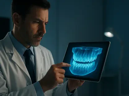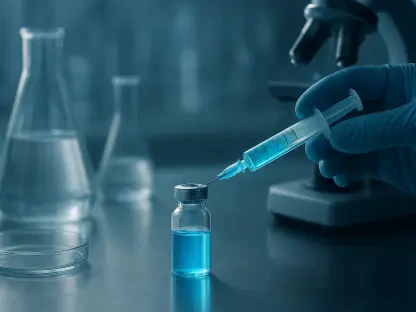

The development of a gelatin methacryloyl hydrogel scaffold provides a biocompatible framework that mimics the natural extracellular matrix for cell growth. This innovation, spearheaded by Professor Jun Liu and a dedicated research team at Sichuan University, marks a pivotal advancement in the field of regenerative medicine during 2026. Their work specifically addresses the multifaceted challenge of periodontal bone loss, a condition that has historically resisted complete restoration through conventional methods. By investigating the molecular intricacies of dental follicle stem cells, or DFSCs, the team has identified a way to bypass the biological limitations imposed by aging and chronic inflammation. This study, featured in the September edition of the International Journal of Oral Science, provides a comprehensive look at how bioengineered systems can restore the regenerative potential of these specialized cells. The research effectively bridges the gap between laboratory molecular biology and practical clinical applications. It offers a beacon of hope for patients suffering from long-term tissue degradation that once seemed irreversible in the face of natural aging processes and the cumulative effects of systemic health decline.
Unlocking the Genetic Key to Cell Rejuvenation
Identifying the Role of IGFBP5 in Cellular Health
The identification of molecular markers that signal cellular decline has been a longstanding goal for scientists seeking to arrest the effects of biological aging. Within the context of dental health, the research team focused on insulin-like growth factor binding protein 5, a regulator that appears to diminish as stem cells lose their vitality. Through rigorous genetic analysis, it was observed that as dental follicle stem cells undergo the natural process of replicative senescence, the expression levels of this specific protein plummet by a factor of nearly seventy. This dramatic reduction serves as a primary driver for the functional decline seen in aging tissues, where the body’s natural reservoir of repair cells becomes increasingly sluggish and unresponsive. By pinpointing this particular protein as a master regulator, the researchers established a clear link between a cell’s genetic output and its physical ability to contribute to the healing of bone and gum tissues. This realization allows for a more targeted approach to therapy, shifting the focus from simply adding more cells to fixing the internal machinery of the cells already present.
Further investigation into the impact of oxidative stress, a common byproduct of chronic inflammation in periodontitis, revealed that environmental factors exacerbate this protein deficiency. Even in relatively young cells, exposure to high levels of reactive oxygen species caused a fivefold decrease in IGFBP5 levels, effectively forcing the cells into a premature state of senescence. This discovery highlighted the fact that the cellular environment is just as critical as chronological age in determining the success of regenerative therapies. When these protein levels are deficient, stem cells lose their ability to survive and migrate toward sites of tissue injury, which explains why many traditional stem cell transplants fail in diseased mouths. The realization that external stress mimics natural aging processes allowed the team to develop a treatment strategy that does not merely replace lost cells but actively restores their internal biochemical balance. By fortifying the cells against oxidative damage, the researchers have created a more resilient population of stem cells capable of thriving in the hostile conditions of an infected or aging oral cavity.
Regulating the Wnt Signaling Pathway
Beyond the mere identification of the protein, the study successfully mapped out the exact biological pathway through which IGFBP5 operates to restore cellular function. The researchers discovered that by artificially overexpressing this protein, they could suppress a specific inhibitory signal known as WNT5B. In the complex world of cell signaling, the Wnt pathway serves as a major regulator of bone homeostasis, but it often becomes imbalanced during illness. The study found that WNT5B acts as a biological “brake” on bone formation, preventing aging stem cells from maturing into the osteoblasts needed to rebuild skeletal structure. By blocking this inhibitory signal, the rejuvenated stem cells regained their full capacity to differentiate and produce mineralized tissue. This IGFBP5-WNT5B axis provides a precise molecular target for future therapies, allowing clinicians to potentially “turn off” the signals that stop bone growth and “turn on” the body’s natural repair mechanisms. This level of precision is essential for developing treatments that are both effective and predictable across different patient populations.
The suppression of the non-canonical WNT5B pathway did not just restart bone formation; it also improved the overall health and metabolic activity of the stem cells. When the IGFBP5 protein was restored to its optimal levels, the downstream effector known as c-Jun was also regulated, leading to a more stable cellular environment. This cascading effect suggests that the rejuvenation process is holistic, affecting multiple layers of the cell’s operational software. By addressing the root cause of the signaling failure, the treatment allows the cells to function as if they were decades younger, effectively resetting their biological clock. This breakthrough represents a significant departure from older methods that relied on heavy doses of growth factors, which often had unpredictable side effects. Instead, this new approach works within the existing framework of the cell’s own genetic instructions, making it a much safer and more harmonious method of tissue engineering. The ability to manipulate these pathways with such accuracy suggests that the future of dentistry will be defined by molecular-level interventions that work in tandem with the body’s natural processes.
Engineering the Future of Bone Regeneration
The Development of the Gel-vHA@oe-DFSC Scaffold
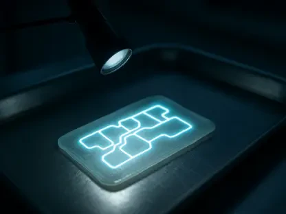
To translate these molecular findings into a practical and usable treatment, the team engineered a sophisticated biomaterial delivery system designed for maximum biocompatibility. This system, a hydrogel scaffold termed Gel-vHA@oe-DFSC, serves as a protective housing for the rejuvenated stem cells during the critical early stages of implantation. By combining a biocompatible gelatin base with vinyl-functionalized nanohydroxyapatite, the scaffold mimics the natural mineralized environment of the bone. The inclusion of nanohydroxyapatite is particularly important because it provides the structural support and essential minerals that encourage stem cells to begin the process of ossification. This engineered environment acts as a bridge, connecting the liquid state of the hydrogel with the solid requirements of the alveolar bone. The resulting material is easy for surgeons to handle and apply to complex periodontal defects, ensuring that the therapeutic cells remain exactly where they are needed to facilitate repair. This structural innovation ensures that the biological benefits of rejuvenated cells are not lost due to poor physical delivery.
The mechanical properties of the scaffold were specifically tuned to match the requirements of the oral cavity, which is subject to constant pressure and movement. The vinyl-functionalized components allow for a stable chemical structure that does not degrade too quickly, giving the stem cells ample time to establish a new tissue matrix. Unlike older generations of hydrogels that were often too soft or lacked the necessary biological cues, this new composite material actively participates in the healing process. It provides a mineral-rich surface that stem cells can grip, which is essential for their migration and eventual differentiation into bone-forming units. The precision of the Gel-vHA@oe-DFSC system represents the next generation of “smart” biomaterials that do more than just fill space; they interact with the body on a cellular level to guide the formation of high-quality tissue. By providing both the “seed” (the rejuvenated cells) and the “soil” (the mineralized scaffold), the researchers have created a comprehensive solution for bone loss that addresses both the biological and physical requirements of regeneration.
Ensuring Cell Survival in Hostile Environments
One of the greatest challenges in regenerative medicine is ensuring that transplanted cells survive the transition into a diseased or inflamed area. The newly developed scaffold acts as a specialized “launchpad,” protecting the modified stem cells from the high levels of oxidative stress and inflammatory cytokines typically found in periodontal tissues. In laboratory settings, the researchers demonstrated that cells housed within this hydrogel maintained their viability even when exposed to harsh chemicals that would normally trigger cell death. This protective effect is crucial because the environment of a patient with periodontitis is often toxic to new cell growth. By creating a micro-environment that shields the stem cells, the scaffold allows them to focus their energy on bone production rather than mere survival. This increased resilience ensures that the therapy remains effective even in patients with active underlying conditions, significantly expanding the pool of candidates who can benefit from such advanced regenerative procedures.
The scaffold also facilitates a steady exchange of nutrients and waste products, which is vital for maintaining the high metabolic activity of rejuvenated cells. Because these cells are engineered to be more active, they require a constant supply of energy and a way to clear out the byproducts of their rapid growth. The porous nature of the gelatin methacryloyl base allows for this flow of fluids, mimicking the natural vascularization found in healthy bone tissue. This ensures that the center of the transplant does not become necrotic, a common failure point for thicker tissue grafts. By optimizing the internal architecture of the scaffold, the team has solved a major hurdle in the scalability of stem cell therapies. The ability of the rejuvenated cells to spread and colonize the surrounding tissue while remaining protected by the hydrogel suggests that this method could be used to treat even large-scale bone defects that were previously considered untreatable. This focus on the long-term survival of the cells is what distinguishes this research from previous attempts at stem cell delivery.
Validating Results Through Clinical Models
Achieving Success in Animal Trials
The efficacy of the rejuvenation method was put to the test in a rat model of periodontitis, yielding results that were both robust and encouraging. Animals treated with the engineered scaffold and rejuvenated cells showed a marked increase in bone mineral density and overall bone volume when compared to those receiving standard treatments. The quantitative data confirmed that the IGFBP5-modified cells were significantly more effective at rebuilding the alveolar bone, which supports the teeth. This was not merely a superficial fix; the newly grown bone exhibited a structural complexity that mirrored healthy, natural tissue. Micro-computed tomography scans revealed that the treated areas had a dense, well-connected network of bone trabeculae, which is essential for withstanding the mechanical stresses of chewing. The success of these trials during 2026 demonstrates that the molecular “reset” of stem cells is a viable path forward for human dentistry, providing a clear proof-of-concept for the next phase of clinical research.
The speed at which the healing occurred was another significant finding of the animal studies. Compared to the control groups, the rats treated with the Gel-vHA@oe-DFSC system reached peak bone density much faster, suggesting that the rejuvenated cells were able to begin the repair process almost immediately upon implantation. This rapid response is vital for preventing further tooth loss and stabilizing the oral environment before more damage can occur. The researchers noted that the integration between the newly formed bone and the original skeletal structure was virtually seamless, with no evidence of the scarring or fibrous tissue that often plagues traditional bone grafts. This high level of integration ensures that the restored bone is functional and capable of supporting dental implants or natural teeth over the long term. By achieving such high-quality results in a living model, the research team has moved one step closer to proving that cellular rejuvenation is the gold standard for future periodontal therapies.
Reducing Inflammation and Improving Tissue Quality
In addition to growing new bone, the treatment significantly improved the health of the surrounding gums and periodontal ligaments, which are often overlooked in traditional bone grafting. The sites treated with the rejuvenated stem cells showed a noticeable reduction in chronic inflammation and a more organized collagen structure, indicating that the cells were influencing the entire local environment. This suggests that the IGFBP5-modified cells do more than just build bone; they act as “bio-modulators” that help reset the entire pocket of infection toward a state of healing and stability. By secreting beneficial signaling molecules, the rejuvenated cells helped to quiet the overactive immune response that typically drives tissue destruction in periodontitis. This dual-action approach—building bone while simultaneously calming the surrounding tissue—is what makes this therapy so revolutionary. It addresses both the symptoms and the underlying biological dysfunction of the disease, leading to a much more stable and long-lasting recovery.
The improved tissue quality was particularly evident in the way the periodontal ligament reattached to the tooth and the newly formed bone. In many cases of advanced gum disease, the ligament is destroyed, making it impossible for the tooth to stay anchored. However, the use of rejuvenated DFSCs appeared to encourage the regrowth of these vital connective fibers, creating a strong and flexible bond. Histological analysis showed that the collagen fibers were oriented in a way that mimicked healthy anatomy, a result that is notoriously difficult to achieve with synthetic materials alone. This finding indicates that the treatment restored the true functional unit of the tooth support system, rather than just the hard tissue. The holistic nature of this repair suggests that patients could see a return to full oral function, including a reduction in tooth mobility and gum recession. The ability to regenerate multiple tissue types simultaneously through a single intervention marks a major milestone in the quest for comprehensive oral rehabilitation.
Broadening the Scope of Stem Cell Therapy
Implications for Age-Related Diseases
While the primary focus of this study was on periodontal health, the discovery of a molecular “reset button” for stem cells has massive implications for general medicine and the treatment of other skeletal disorders. Cellular senescence is a universal biological problem that contributes to a wide range of age-related conditions, including osteoporosis and chronic non-healing fractures. The ability to identify and manipulate a master regulator like IGFBP5 offers a potential blueprint for rejuvenating various types of adult stem cells found throughout the human body. For instance, the same principles applied to dental follicle stem cells could potentially be adapted for bone marrow-derived stem cells used in orthopedic surgeries. By pre-treating these cells to boost their IGFBP5 levels, surgeons could significantly improve the success rates of joint replacements and spinal fusions, particularly in elderly patients whose natural healing capacity is diminished. This research opens the door to a new era of age-defying medicine where the limitations of the aging body are no longer a barrier to recovery.
Beyond bone health, the management of the Wnt signaling pathway through IGFBP5 could have applications in treating cardiovascular disease and other inflammatory conditions. Since chronic inflammation is a common thread in many systemic diseases, the bio-modulatory effects observed in the periodontal tissues might be replicable in other organs. If researchers can harness the anti-inflammatory and regenerative properties of these rejuvenated cells, they might develop new therapies for repairing damaged heart tissue after a myocardial infarction or treating degenerative joint diseases like arthritis. The study provides a valuable framework for understanding how to balance complex signaling networks to promote healing rather than decay. As the global population continues to age, the demand for these types of precision regenerative therapies will only increase. The Sichuan University study serves as a foundational text for this emerging field, proving that we can fundamentally change how the body responds to the passage of time by intervening at the genetic and molecular levels.
Navigating the Path to Human Application
The transition from animal models to human clinical application represents the final and most critical hurdle for this technology. Future efforts must focus on optimizing the dosage of IGFBP5 and ensuring that the hydrogel scaffold can be manufactured consistently on a commercial scale. Researchers previously determined that while the rat model provided excellent proof-of-concept, the biological complexity of the human oral microbiome might require additional modifications to the delivery system. For example, the scaffold may need to be enhanced with antimicrobial properties to ensure that the rejuvenated cells are not overwhelmed by local bacteria during the initial healing phase. Furthermore, clinical trials will need to assess how cells harvested from older human patients with varying health backgrounds respond to the rejuvenation process. This phase of development was initiated to ensure that the treatment is as safe as it is effective, with long-term monitoring of patient outcomes being a top priority for the scientific community.
The success of these initial studies paved the way for a more personalized approach to regenerative dentistry, where a patient’s own “tired” cells were harvested, rejuvenated in a lab, and then returned to their body to fix specific defects. Actionable next steps included the development of standardized protocols for the extraction and modification of stem cells to minimize the time between diagnosis and treatment. Industry leaders then looked toward integrating this technology into routine clinical practice, aiming to make it as accessible as traditional bone grafting. The insights gained from this research provided a clear roadmap for the future of skeletal medicine, emphasizing the need for a deep understanding of cellular pathways. By focusing on the functional optimization of cells rather than just their quantity, the medical community moved toward a more sophisticated model of care. This work ultimately laid the groundwork for a future where aging and disease were no longer permanent obstacles to maintaining a healthy and functional skeletal system.