
Pedicle screw placement requires sub-millimeter accuracy because a deviation of only two millimeters can cause permanent nerve damage or severe vascular injury. In the modern surgical suite, the challenge is not just the operation itself but balancing the necessity of precise visualization with the dangers of prolonged radiation exposure. Surgeons traditionally depend on intraoperative X-rays or fluoroscopy to verify the position of their instruments, yet this repetitive imaging accumulates health risks over a career. As the medical community moves toward safer alternatives, the integration of non-ionizing modalities has become a priority. A pioneering research effort at Beijing Shijitan Hospital has successfully addressed this by combining the real-time capabilities of ultrasound with high-resolution 3D cone-beam computed tomography. This multimodal approach allows for a navigation map that is both incredibly accurate and fundamentally safer for everyone in the room. By achieving a precision level of approximately one millimeter, this technology bridges a critical gap in contemporary spinal intervention.
The Challenge: Static Maps vs. Dynamic Patient Anatomy
The primary hurdle in modern spinal navigation is often referred to as the “stale map” problem. While cone-beam computed tomography provides a high-resolution 3D view of the bone, it remains a static image captured at a single point in time. During the course of a procedure, the patient’s body may shift slightly, or the physical manipulation of the vertebrae might change the actual alignment of the spine. When these shifts occur, the initial CT scan no longer represents the patient’s physical reality on the operating table, which often forces surgical teams to pause and take additional X-rays to recalibrate their navigation systems. This cycle not only increases the procedure’s duration but also subjects the patient to further radiation. Solving this requires a dynamic imaging source that can update the surgeon’s view without the drawbacks of ionizing rays. The objective is to keep the surgical map synchronized with the physical body in real time, even as conditions shift.
Ultrasound serves as a promising alternative in this context because it is portable, provides live updates, and involves no radiation whatsoever. However, ultrasound technology has historically been difficult to utilize effectively during bone surgery. Sound waves do not penetrate hard surfaces well, which typically results in noisy or grainy images that only depict the bone’s outer shell rather than its internal structure. Furthermore, the presence of soft tissue and air can create artifacts that obscure the very landmarks surgeons need to see. The technical challenge lies in the complex task of stitching these grainy, partial ultrasound surfaces onto the high-quality, complete anatomical models provided by a pre-operative or early-operative CT scan. If the system cannot perfectly align the noisy live feed with the clear static model, the navigation becomes untrustworthy. Bridging this data gap requires advanced mathematical algorithms capable of interpreting signals and mapping them onto a rigid frame.
System Architecture: Integrating Robotics and Calibration
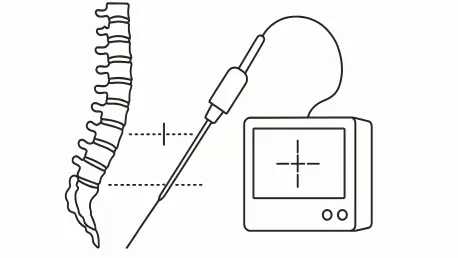
To overcome the limitations of manual imaging, the researchers developed a sophisticated navigation system that integrates an ultrasound machine, a mobile C-arm for CT scans, and an optical tracking system. The inclusion of an optical system is vital, as it uses infrared cameras to monitor the physical position of surgical tools and imaging probes in three-dimensional space with extreme precision. To ensure the highest level of consistency during data acquisition, the team avoided manual scanning, which can be prone to human error and variable pressure. Instead, they employed a robot-controlled arm to move the ultrasound probe across the patient’s anatomy at a steady, controlled speed of five millimeters per second. This robotic precision ensures that the resulting images are uniform and free from the jitters or inconsistencies that occur when a human hand handles the transducer. By automating the sweep, the system generates a high-quality dataset that serves as the foundation.
The experimental phase utilized a lumbar spine phantom, which is a realistic model of the L1 through L4 vertebrae designed to mimic human bone density. To create an ideal imaging environment for these initial tests, the researchers submerged the phantom in water. This specific setup was intentional, as water eliminates the air-tissue interference that usually degrades ultrasound quality in a living clinical setting. By removing these biological variables, the team was able to isolate and measure the raw accuracy of their alignment algorithms. This allowed them to prove that the underlying software and robotic hardware could theoretically reach sub-millimeter precision before introducing the complexities of human flesh. This controlled environment provided the necessary baseline to confirm that any errors observed were the result of the technology itself rather than external noise. This methodology ensured that the fusion process was scientifically sound and capable of handling data processing.
Image Registration: The Three-Stage Fusion Pipeline
Before any meaningful fusion of ultrasound and CT data could occur, the system required an incredibly precise calibration process to establish a common language between the devices. This involved teaching the computer exactly how the pixels in a two-dimensional ultrasound image corresponded to the physical location of the probe in the three-dimensional operating environment. To achieve this, the team used a specialized calibration phantom containing N-shaped wires. By imaging these wires from eighty different angles, the system could calculate the exact coordinate transformation needed to turn a flat image into a spatial map. The level of detail achieved during this phase was remarkable, resulting in an ultrasound image scale of just 0.16 millimeters per pixel. This step is the most critical part of the entire workflow, as any error in the initial calibration would be amplified throughout the surgery, leading to dangerous deviations in screw placement. Without this groundwork, navigation would fail.
The core of the team’s success was a three-step registration process designed to align the live ultrasound data with the high-resolution CT model. The first stage involved a similarity transform, which acted as a rough digital alignment to bring the two different types of images into the same general neighborhood in the software’s coordinate system. Once the images were roughly aligned, the system moved into the second stage, utilizing an Iterative Closest Point algorithm. This mathematical tool works by identifying the nearest points between the ultrasound surface and the CT model and snapping them together to create a rigid alignment. While this stage significantly improved the accuracy, it still treated the bone surfaces as rigid objects that might not account for subtle variations or noise in the ultrasound signal. Despite these improvements, the rigid alignment alone was not quite precise enough to meet the strict sub-millimeter safety standards required for the most delicate parts.
Precision Benchmarks: Breaking the Millimeter Barrier
The final and most innovative step of the process involved the application of Thin-Plate Spline deformation. This mathematical tool is a form of non-rigid refinement that treats the ultrasound image as if it were a flexible sheet that can be slightly warped or bent to perfectly contour to the detailed CT model. This step is crucial because it accounts for the small errors caused by soft-tissue interference or the limited surface visibility that is inherent in ultrasound technology. By allowing for these minute adjustments, the system can stretch the noisy ultrasound data to find the best possible fit against the gold standard CT anatomy. This transition from a rigid to a non-rigid alignment model is what allowed the researchers to break through previous accuracy barriers. The Thin-Plate Spline approach ensures that even if the ultrasound data is slightly distorted by environmental factors, the final fused map remains anchored to the true anatomical structure of the patient’s spine.
When the researchers evaluated the accuracy using only the rigid Iterative Closest Point step, the error margin sat at nearly 3 millimeters. In the world of spinal surgery, a three-millimeter deviation is often considered the upper limit of safety, as it leaves almost no room for error when navigating near the spinal cord. However, once the Thin-Plate Spline refinement was applied to the data, the target registration error dropped significantly to an average of just 1.26 millimeters. In addition to average accuracy, the researchers measured the worst-case scenario using the Hausdorff distance, which identifies the maximum misalignment between surfaces. After the final non-rigid processing stage, the most misaligned parts of the fused model were accurate to within 1.54 millimeters. This means that the entire digital map used by the surgeon is roughly as accurate as the thickness of a typical coin, providing a level of precision that allows for much more aggressive and effective stabilization.
Clinical Implementation: Efficiency and Future Considerations
For any new technology to be adopted by the medical community, it had to be efficient enough to fit into the fast-paced and high-pressure environment of a modern operating suite. The study found that the entire workflow—including the robotic scan of the anatomy and the computer’s subsequent data processing—took less than three minutes to complete. Specifically, the robotic sweep across a 170-millimeter section of the spine took only 34 seconds, while the complex fusion calculations required approximately 144 seconds. This rapid turnaround ensured that the navigation setup did not unnecessarily prolong the time a patient spent under anesthesia, which was a key factor in reducing surgical complications. By streamlining the registration process, the system offered a practical solution that could be integrated into existing hospital workflows without requiring significant changes to the way surgeons and nurses prepared for a surgical case throughout the current 2026 period.
Looking ahead, the researchers planned to transition into cadaveric studies to better simulate the density and complexity of human tissue. These upcoming trials were considered essential for refining the Thin-Plate Spline algorithms to handle the non-linear distortions caused by soft tissue layers. The ultimate goal was to create a commercial-grade navigation platform that provided a simplified experience for hospitals. By proving that millimeter-level precision was attainable through robotic control and advanced image fusion, the team established a viable blueprint for the next generation of spinal care. This research represented a significant shift in the industry, moving away from a total reliance on harmful radiation toward a more intelligent, multi-modal approach. For the thousands of patients who underwent spinal stabilization, these advancements suggested a future where operations were not only more successful but were performed with a significantly higher safety standard.