
By mimicking the bioelectric environment of living bone, this new composite material encourages the body to repair itself using internal biomechanical forces. Orthopedic surgeons have long encountered severe difficulties when treating large segmental bone defects, which are substantial gaps in skeletal structure that cannot heal on their own due to their sheer size. These injuries frequently stem from high-energy trauma, the excision of aggressive tumors, or complex congenital conditions that disrupt the natural regenerative capacity of human bone. While traditional methods like inert metal implants or synthetic ceramics offer necessary structural support, they often fail to integrate biologically, leading to issues such as stress shielding where the surrounding bone weakens because it is no longer carrying weight. Furthermore, autologous bone grafts, though considered a standard, are limited by the availability of donor tissue and the risk of complications at the surgical site. This environment necessitated a transition toward more dynamic, bio-responsive solutions that can actively participate in the healing process rather than just providing a static bridge.
Materials Science: Architectural Design
Integrating Advanced Nanomaterials and Biocompatible Scaffolds
The foundational innovation of this technology lies in a composite material designated as BPNS@rhCOL1MA, which strategically combines black phosphorus nanosheets with a highly specialized collagen matrix. Black phosphorus has emerged as a revolutionary two-dimensional nanomaterial in bone tissue engineering because it possesses inherent piezoelectric properties, meaning it generates an electrical charge in response to physical stress. This phenomenon closely mirrors the natural bioelectric signals produced by healthy bone when it is subjected to mechanical loads during daily activity. Beyond its electrical capabilities, black phosphorus is exceptionally efficient at converting specific wavelengths of light into local therapeutic heat. Crucially, as the material performs its function, it eventually biodegrades into phosphate ions, which are the fundamental building blocks used by the human body to synthesize new hydroxyapatite for bone mineralization. This dual-purpose role as both a stimulator and a raw material source makes it an ideal candidate for regenerative orthopedic applications.
To translate these nanomaterial benefits into a clinical setting, the research team successfully encapsulated the nanosheets within a hydrogel composed of recombinant humanized type I collagen. This choice of scaffold represents a significant leap forward from traditional animal-derived collagen products, which have historically carried risks of triggering unwanted immune responses or transmitting pathogens. By producing this collagen through microbial fermentation, scientists have created a material that is nearly identical to human tissue, ensuring high biocompatibility and a reduced risk of rejection. The collagen is modified with methacrylate groups, which allows the liquid solution to be cross-linked into a stable three-dimensional network upon exposure to a specific light source. This ability to exist in a liquid state before hardening allows the hydrogel to be injected into irregular and complex bone voids, where it molds itself to the unique shape of the injury before setting into a supportive, bioactive matrix that protects the delicate black phosphorus from premature oxidation.
Precision Engineering: Tissue Integration
Sophisticated imaging and materials characterization have confirmed that the resulting hydrogel maintains an ideal architectural environment for cellular growth. Using transmission electron microscopy and atomic force microscopy, the development team verified that the black phosphorus remained in its high-performance crystalline nanosheet form throughout the fabrication process. The internal structure of the hydrogel features a highly porous and interconnected network, which is vital for the long-term survival of the implant. This biomimetic “sponge-like” design facilitates the continuous flow of oxygen and essential nutrients to the cells residing deep within the scaffold while simultaneously allowing for the removal of metabolic waste products. Furthermore, this open architecture provides the necessary physical space for angiogenesis, the process by which new blood vessels infiltrate the repair site to provide the vascular support system required for permanent, living bone tissue to replace the temporary hydrogel.
The engineering of this material also allows for the precise adjustment of its mechanical properties to suit different clinical needs. By varying the concentration of the black phosphorus nanosheets, researchers can fine-tune the storage modulus, or “stiffness,” of the gel. This ensures that the material is robust enough to resist being displaced by internal physiological pressures while remaining flexible enough to transition energy effectively. Zeta potential measurements indicated a stable negative charge on the surface of the nanosheets, a critical factor that prevents the particles from clumping together and ensures a uniform distribution of the piezoelectric and photothermal effects throughout the entire defect area. Because the integration process does not damage the molecular integrity of the collagen, the two components work in perfect synergy, creating a stable platform that supports both the structural and biological requirements of the healing site in high-stress environments.
Accelerated Healing: Mechanics and Implementation
Harvesting Energy: Physiological Movements
The most distinctive characteristic of this injectable system is its ability to harvest energy from the body’s own rhythmic movements, specifically the act of breathing. When the hydrogel is implanted in areas such as the rib cage or near the diaphragm, the constant mechanical strain of respiration triggers the piezoelectric effect within the black phosphorus nanosheets. This results in the generation of a stable voltage output that is significantly higher than the bioelectric fields naturally produced by living bone tissue. This means that the simple, passive act of breathing serves as a continuous power source for the healing process, eliminating the need for bulky external electrical stimulation devices or invasive battery-powered implants. This constant bioelectric field acts as a persistent signal to the surrounding biological environment, informing the body that active repair and tissue reinforcement are required in that specific location.
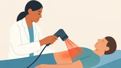
In addition to this passive energy harvesting, the material incorporates an active external trigger through the use of near-infrared (NIR) light therapy. When the treatment area is exposed to 808-nanometer NIR light, the black phosphorus nanosheets within the hydrogel efficiently convert that light energy into localized heat. This process is carefully controlled to reach a range of “mild hyperthermia,” typically between 104 and 107 degrees Fahrenheit. This specific temperature range has been shown to enhance local blood circulation and increase the metabolic rate of cells without causing any thermal damage to the surrounding healthy tissue. The system demonstrated remarkable photothermal stability, maintaining its conversion efficiency through repeated cycles of activation. This reliability allows for a treatment regimen where a patient could receive short, non-invasive light treatments daily, complementing the continuous piezoelectric stimulation provided by their own breathing to maximize the rate of bone regeneration.
Driving Stem Cell: Transformation and Bone Formation
The biological effectiveness of the dual-stimulus hydrogel was confirmed through extensive studies involving bone marrow mesenchymal stem cells, which are the body’s primary agents for skeletal repair. When these cells were cultured on the hydrogel and exposed to both the piezoelectric signals and the light-triggered heat, they exhibited significantly higher rates of proliferation and survival compared to those on standard scaffolds. The combined physical signals serve as a powerful set of instructions, essentially “programming” the stem cells to begin the complex process of differentiation. Molecular analysis of these cells revealed a sharp increase in the expression of key osteogenic markers, such as alkaline phosphatase and osteocalcin, which are definitive indicators that the stem cells are successfully maturing into functional bone-forming cells. This transition is essential for moving beyond the initial inflammatory response and toward the actual construction of new mineralized tissue.
To gain a deeper understanding of these cellular mechanisms, researchers utilized transcriptomic sequencing to observe how thousands of genes responded to the hydrogel’s stimuli. The data showed that nearly 4,000 genes were upregulated, particularly those involved in pathways responsible for sensing mechanical forces and initiating the formation of new blood vessels. The team identified that the mild heat increases the fluidity of cell membranes, while the piezoelectric charges activate specific mechanical sensors like the Piezo1 protein. These physical interactions then trigger internal signaling cascades, such as the MAPK/ERK and BMP/Smad pathways, which carry the instructions from the cell surface directly to the nucleus to initiate bone production. This comprehensive “stimulus-to-formation” axis provides a clear scientific explanation for how the hydrogel manages to coordinate such a rapid and effective healing response by speaking the same physical and chemical language as the body’s own cells.
Rapid Regeneration: Success in Living Models
The definitive proof of this technology’s potential was demonstrated through successful trials in animal models, where critical-sized defects in the ribs were healed with unprecedented speed. In these experiments, the natural respiratory movement of the rib cage provided the continuous mechanical stress needed to activate the piezoelectric properties of the black phosphorus. While control groups with no treatment or standard collagen gels showed incomplete healing and persistent gaps, the rabbits treated with the dual-stimulus hydrogel achieved full bone bridging in just four weeks. This represents a massive improvement over the typical three-month healing period required for such significant injuries. The regenerated bone was not merely a superficial bridge; it displayed high mineral density and mechanical strength that were virtually indistinguishable from the animal’s original, healthy bone tissue, proving that the accelerated timeline did not compromise the quality of the repair.
Histological examinations during the study confirmed that the hydrogel promoted a natural biological pathway known as endochondral ossification, where the body first established a cartilage-like template before rapidly replacing it with mature, vascularized bone. Throughout the observation period, the biodegradable components of the hydrogel were processed safely by the animals, with no evidence of systemic toxicity or damage to internal organs. This established a clear safety profile for the materials involved, suggesting a straightforward path toward clinical trials for human application. As the research reached its conclusion, the results supported a future where complex reconstructive surgeries could be simplified through a single injection of a smart material. These findings indicated that by harnessing the body’s internal energy and augmenting it with targeted external stimuli, the medical community could successfully reduce recovery times from several months to a matter of weeks, fundamentally changing the prognosis for patients with severe skeletal trauma.