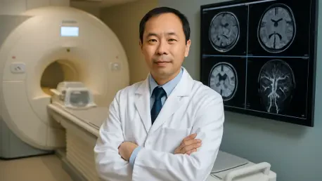

Reducing MRI scan times to under four minutes has proven essential for minimizing motion artifacts and improving the comfort of patients experiencing acute vascular distress. At the University of California, San Francisco, Dr. Zhaoyang Fan is leveraging his background in both radiology and engineering to push these boundaries further than ever before. As the co-chief of Cardiac and Vascular Imaging Research, he operates at the vital intersection where complex technical development meets real-world clinical application. His work is not merely about producing clearer images; it is about redefining how physicians understand the structural integrity of the human vascular system. By focusing on the subtle nuances of arterial health, Dr. Fan provides clinicians with the tools necessary to predict and prevent catastrophic events like strokes. This interdisciplinary approach ensures that the engineering innovations created in the laboratory are directly translated into diagnostic protocols that can be implemented in high-pressure medical environments.
The Science: Advancing Precision Through Vessel Wall Imaging
A primary cornerstone of this research is the advancement of Vessel Wall Imaging, a technique frequently referred to as “black blood” imaging. While traditional magnetic resonance imaging techniques typically focus on the flow of blood through an artery, the methodology developed here suppresses the signal from the blood itself to highlight the architecture of the vessel wall. This specific effect allows clinicians to identify atherosclerotic plaques that are at a significantly high risk of rupturing, which is a detail often missed by conventional scans. By visualizing these hidden dangers with high-resolution clarity, doctors are now able to monitor changes over time, providing a longitudinal perspective that is far more useful for long-term patient care than a single, static snapshot. This shift in focus from the void of the artery to the physical composition of its boundaries represents a fundamental change in how vascular disease is managed, moving from reactive treatment to proactive risk assessment.
The integration of speed and quantitative accuracy has been the most significant hurdle in medical imaging, often requiring a trade-off between image quality and scan duration. To address this, the laboratory has pioneered new acquisition sequences that have slashed scan times to under four minutes, a breakthrough that significantly reduces motion blur caused by patient movement. Beyond simple speed, the introduction of automated software has shifted the diagnostic process from subjective observation to quantitative measurement. Instead of relying solely on a radiologist’s visual intuition, these tools provide objective data, allowing for precise tracking of whether a patient’s treatment plan is actually working. These advancements ensure that the data collected is not only faster to acquire but also more reliable for longitudinal studies. This transition toward data-driven diagnostics allows for a more granular understanding of how individual patients respond to various therapies over weeks or months of observation.
Global Implementation: Scaling Diagnostic Impact for Stroke Care
In the high-stakes environment of acute stroke management, speed and accuracy are the most vital factors in determining a patient’s eventual outcome. Development is currently focused on a comprehensive “all-in-one” MRI protocol that examines the heart, neck, and brain in a single session lasting less than ten minutes. This integrated approach helps physicians quickly determine if a stroke was caused by a local blockage in the brain or an embolism traveling from the heart. By streamlining this diagnostic window, healthcare providers can implement the correct treatment protocol faster, significantly reducing the risk of a second, more damaging stroke. This holistic view of the vascular system provides a level of diagnostic certainty that was previously unattainable in such a short timeframe. The protocol effectively removes the guesswork from the initial triage phase, ensuring that the source of the vascular event is identified with precision before the appropriate neurological intervention begins.
Scientific focus was also shaped by a sense of social responsibility regarding global health trends, particularly in East Asia where carotid plaque remained a leading cause of stroke. The team recognized that perfecting techniques to identify vulnerable patients before a major vascular event occurred served as a vital tool for improving public health outcomes on a global scale. This effort culminated in the realization that advanced imaging technology had to reach the populations most at risk to be truly effective. The success of these breakthroughs was ultimately rooted in a deep commitment to collaborative science, which utilized virtual platforms to connect experts across various institutions. These partnerships proved that integrating diverse expertise could solve complex medical challenges. The transition from laboratory innovation to life-saving bedside application was completed through sustained dedication to engineering excellence. Future research trajectories began to focus on AI-driven predictive modeling to further refine stroke prevention strategies.