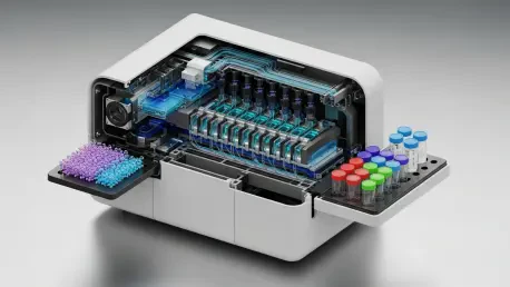

Hospitals needed faster answers while disease often hid in the subtle give, drag, and spring of living cells as they squeezed through microchannels and flowed past sensors that could now read mechanics at scale. That tension between urgency and nuance defined the promise of a new microfluidic “mechanophenotyping cytometer,” built to read cellular stiffness alongside conventional fluorescence signals. The device, developed through an academic–standards agency partnership, set out to transform a slow, expert-only measurement into a high-throughput, reproducible workflow.
This FAQ mapped the big questions behind the technology: why stiffness mattered, how a flow-based cytometer could infer it without poking cells, and where such data might change clinical decisions. Readers could expect clear explanations, relevant examples from cancer and blood disorders, and a balanced view of practical hurdles such as calibration, accuracy, and standardization.
Key Questions or Key Topics Section
Why Does Cell Stiffness Matter for Health and Disease?
For decades, researchers observed that tumor cells tend to soften as they acquire invasive traits, while diseases like malaria and sickle cell disease make red blood cells stiffer. Those mechanical shifts reflect underlying cytoskeletal remodeling and membrane changes, which can parallel disease onset, progression, or treatment response. Mechanics, in other words, carried biological meaning that traditional molecular markers sometimes missed.
Stiffness also varied in neurodegenerative, cardiovascular, and chronic inflammatory conditions, making mechanics a cross-cutting biomarker. When sampled across thousands of cells, mechanical phenotypes could help separate healthy from diseased populations, uncover heterogeneity, and reveal early transitions that influence prognosis or therapy selection.
What Held Mechanophenotyping Back Until Now?
The gold standard, atomic force microscopy, indented cells one at a time with a microscopic probe. While exquisitely sensitive, AFM was slow, operator-dependent, and limited by where and how the probe contacted the cell. Processing large, heterogeneous clinical samples within practical timelines became difficult, and inconsistent protocols made cross-lab comparisons a challenge.
Those constraints made mechanophenotyping rare in clinics despite its promise. A scalable, automated, and standardized method was needed to move mechanics from boutique measurements into routine assays that could compete with flow cytometry on speed and reliability.
How Can a Cytometer Infer Stiffness Without Touching the Cell?
The new device flipped the script by using time-of-flight through a microfluidic channel as a proxy for stiffness. In laminar flow, fluid moved fastest in the center and slower near the walls; softer cells tended to migrate toward the centerline and travel faster, while stiffer cells remained near slower streamlines. By correlating each cell’s travel time between checkpoints with its size, the system inferred relative stiffness.
Fluorescence signals, already standard in cytometry, provided size estimates and phenotypic tags. Multiple measurement regions in series supplied repeated reads on the same cell, yielding built-in error bars and exposing both technical and biological variability—key ingredients for trustworthy diagnostics.
How Fast and Reliable Is This Approach Compared With AFM?
Where AFM might reach roughly one cell every 30 seconds in skilled hands, the mechanophenotyping cytometer measured about 60 to 100 cells per second during tests. The hardware and physics supported rates in the hundreds or thousands per second, opening the door to population-scale analyses that capture rare cells and subtle shifts.
Calibration underpinned reliability. Polymer-based cell mimics with known sizes and stiffnesses produced clear, theory-aligned correlations between size, speed, and inferred modulus. Repeated checkpoints quantified uncertainty, and the tight alignment with predictions contrasted with prior methods that often delivered variable or sample-prep–sensitive results.
Can This Integrate With Existing Lab Workflows?
Yes. Because the platform leveraged fluorescence detection used by mainstream cytometers, it slotted into familiar optics and electronics. Sample preparation echoed standard cytometry steps, reducing the learning curve and making it easier to pilot in research cores and translational labs.
Moreover, coupling mechanics with multiparametric fluorescence meant stiffness could be read alongside markers of lineage, activation, or mutation. That combination strengthened specificity: a soft, invasive phenotype could be linked to a known tumor subpopulation, while a stiff red cell fraction could be tied to hematologic genotypes or infection.
What Are the Most Promising Applications?
Immediate targets included cancers and hemoglobinopathies, where mechanical differences are pronounced and clinically relevant. In oncology, mechanics could assist with risk stratification, minimal residual disease monitoring, or early response assessment. In blood disorders, shifts in stiffness distributions might flag disease severity or therapy effects.
Beyond those, inflammatory and neurodegenerative diseases often carry subtler mechanical changes. High-throughput reads could uncover signatures that complement molecular assays, helping refine diagnoses or identify treatment windows that molecular panels alone might miss.
What Caveats and Open Questions Remain?
Biology is messy: stiffness varies with cell cycle, activation state, and microenvironment. Translating time-of-flight to modulus in mixed clinical samples required careful gating, reference standards, and model refinement. Head-to-head comparisons against AFM and other deformability cytometers across diverse cell types would further anchor accuracy.
Standardization also mattered. Robust calibration beads or gels, harmonized protocols, and validated uncertainty estimates were prerequisites for regulated use. Most importantly, clinical studies needed to show that mechanical readouts improved diagnostic accuracy, prognosis, or monitoring beyond current best practices.
Summary or Recap
Cell mechanics captured disease-relevant information that molecular markers did not always reveal, but traditional tools were too slow and variable for routine use. A new microfluidic cytometer answered that challenge by inferring stiffness from how cells moved through controlled flows, pairing time-of-flight with fluorescence-based size.
Early results showed high throughput, repeat measurements for in-line error estimates, and strong agreement with theory using calibrated cell mimics. Integration with standard cytometry workflows enabled combined mechanical and molecular profiling, setting the stage for applications in cancer, blood disorders, and potentially inflammatory and neurodegenerative disease.
For deeper reading, explore peer-reviewed work on mechanobiology, microfluidic deformability assays, and standards from measurement institutes that address calibration and reproducibility. Reports in journals focused on lab-on-a-chip technologies provided additional technical context and performance data.
Conclusion or Final Thoughts
This FAQ closed on a practical note: the path forward favored studies that benchmarked accuracy, defined calibration materials, and demonstrated clinical utility in real patient cohorts. Teams that paired mechanical phenotypes with existing markers and clear uncertainty estimates were positioned to make rapid progress.
Adoption also depended on interoperability with current cytometers, standardized software for time-of-flight analysis, and clear guidelines for sample handling. If those steps were met, mechanophenotyping by flow stood to become a credible companion diagnostic—flagging disease presence, refining risk, and tracking treatment—while keeping speed and reproducibility at the forefront.