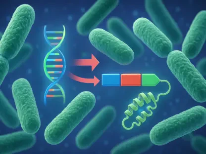

Hyaluronic acid-decorated carriers target the CD44 receptor, which is frequently overexpressed in bladder tumors, to improve the specificity of localized therapy. The staggering incidence of urologic malignancies, which currently accounts for more than 2.6 million new cases and 700,000 deaths annually worldwide, has necessitated a fundamental shift in the way clinicians and bioengineers approach drug delivery for the bladder, prostate, and kidney. While traditional treatments such as surgery, radiotherapy, and systemic chemotherapy remain the pillars of care, they are often hindered by high rates of recurrence and significant systemic toxicity that limits their long-term viability. In 2026, the movement toward an anatomy-guided framework for nanomedicine represents a sophisticated alternative to the conventional “one-size-fits-all” drug administration model. This strategic paradigm posits that the unique physical and structural characteristics of the organ itself must dictate the design of the nanocarrier. By categorizing these organs into distinct anatomical profiles—hollow organs like the bladder, solid glandular structures like the prostate, and highly vascularized parenchymal tissues like the kidney—researchers can tailor the route of administration to bypass physiological barriers. This ensures that therapeutic agents are delivered exactly where they are most effective, maximizing local drug concentrations while shielding the rest of the body from unnecessary exposure.
Bladder Cancer: Overcoming Physiological Barriers
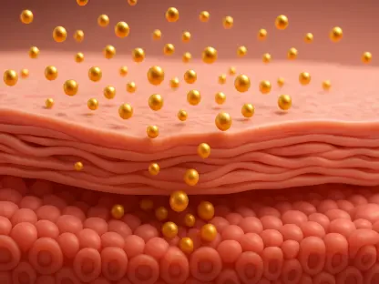
Within the realm of bladder cancer treatment, the shift toward localized intravesical therapy has faced significant hurdles due to the organ’s natural physiology and defensive mechanisms. The primary challenge remains the constant turnover and dilution caused by urine production, combined with regular voiding that flushes therapeutic agents out of the system before they can be effectively absorbed by the malignant tissue. Furthermore, the bladder is lined with a highly resistant glycosaminoglycan layer that acts as a physical shield against the adhesion of foreign substances, including many standard chemotherapeutic drugs. To overcome these persistent obstacles, scientists have turned to bio-inspired chemistry, specifically focusing on the development of mucoadhesive nanoparticles that can cling to the wet mucosal wall. Taking inspiration from the adhesive proteins used by marine mussels, researchers have engineered catechol-functionalized alginate particles and thiolated silica nanoparticles that form robust bonds with the bladder lining. Additionally, cationic polypeptide nanogels utilize electrostatic attraction to bind with the negatively charged surface of the mucosa. This approach ensures that the medication remains in direct contact with the tumor for extended periods, increasing the duration of drug exposure and improving the overall therapeutic index.
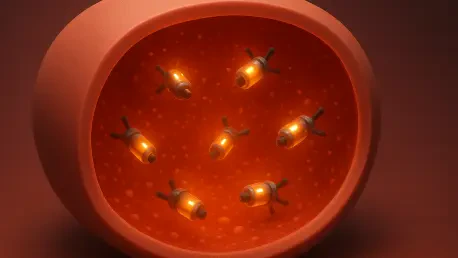
Beyond passive adhesion, the integration of active delivery systems is revolutionizing how we penetrate the dense protective layers of the bladder. One of the most significant breakthroughs in 2026 involves the use of urease-powered nanomotors, which are microscopic carriers that utilize the urea naturally present in urine as a source of fuel for autonomous propulsion. These nanomotors can effectively swim through the mucus layer, reaching the underlying tumor tissue with much higher efficiency than static particles. Other advanced systems leverage near-infrared light-driven motors to achieve mechanical penetration, allowing for high-precision targeting of the tumor microenvironment. By combining these active movement capabilities with stimuli-responsive properties, such as pH or enzyme triggers, these carriers can release their payload only when they have successfully breached the tumor’s defenses. This level of control is essential for treating non-muscle-invasive bladder cancer, where the goal is to eliminate malignant cells without causing widespread inflammation of the bladder wall. The synergy between molecular targeting and mechanical propulsion provides a dual-layered approach to therapy, ensuring that high concentrations of the drug reach even the deepest recesses of the tumor while minimizing the frequency of treatments required for the patient.
Prostate Oncology: Navigating Structural Resistance
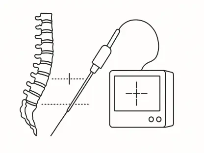
The prostate presents a fundamentally different set of anatomical challenges compared to the bladder, as it is a solid, glandular organ situated deep within the pelvic cavity without a natural lumen for easy drug instillation. Standard systemic therapies often struggle to achieve therapeutic levels within the prostate due to the organ’s dense fibromuscular stroma and high interstitial fluid pressure, which act as formidable physical barriers to diffusion. In response, modern urologic care has increasingly turned to focal therapy, which involves the direct injection of nanomedicines into the prostate tissue. However, simple needle injection is often insufficient, as the drug may leak along the needle track or fail to spread evenly throughout the heterogeneous tumor environment. To address this, researchers are employing mesoporous silica nanoparticles as a versatile delivery platform that can encapsulate high doses of chemotherapy while protecting them from premature degradation. These silica matrices are engineered to release their cargo in a controlled, sustained manner, maintaining high local concentrations within the gland while preventing the systemic side effects, such as neuropathy and bone marrow suppression, that often plague patients undergoing traditional chemotherapy.
Precision in prostate cancer management is further refined through the use of molecular ligands that target the prostate-specific membrane antigen, which is frequently overexpressed in malignant prostate cells. By conjugating these ligands to the surface of nanoparticles, clinicians can achieve a level of cellular-level selectivity that complements the macroscopic localization provided by focal injection. This is particularly vital in the context of multifocal disease, where several small tumors may be scattered throughout the gland and could be missed by standard needle placement. The integration of image-guided technology, such as MRI-visible gold or gadolinium nanoparticles, allows for the real-time monitoring of drug distribution within the organ, ensuring that the therapeutic depot is accurately placed and remains stable. This dual-targeting logic—combining physical placement with molecular specificity—enables the treatment of both the primary tumor mass and any surrounding micro-metastases within the gland. Furthermore, these injectable systems can be loaded with radiosensitizers to enhance the efficacy of focal radiotherapy, creating a synergistic effect that maximizes cancer cell death while sparing the surrounding healthy tissues of the rectum and urethra from collateral damage.
Renal Malignancies: Advancing Treatment Precision
Kidney tumors represent perhaps the most complex target for localized delivery, as the organ lacks a lumen and is characterized by a highly vascularized structure that makes direct tissue penetration risky. In 2026, interventional oncology has bridged this gap by utilizing the renal arterial system as a conduit for anatomy-guided nanomedicine. For patients with clear cell renal cell carcinoma, which typically displays a hypervascular profile, researchers have developed drug-eluting beads and microspheres specifically designed for chemoembolization. This technique involves the precise delivery of particles into the blood vessels feeding the tumor, where they serve a dual purpose: they physically block the blood supply to starve the tumor of oxygen and nutrients while simultaneously releasing high concentrations of chemotherapeutic agents directly into the malignant tissue. The narrow margin for error in this procedure requires nanoparticles with highly controlled size distributions to prevent them from escaping into the general circulation or damaging healthy nephrons, which could lead to kidney failure. By localizing the therapy within the vascular architecture of the tumor, clinicians can achieve a dramatic reduction in tumor volume while preserving the overall renal function, which is a critical consideration for patients with underlying chronic kidney disease.
For cases where surgical intervention is necessary, the use of biomimetic hydrogels has emerged as a powerful strategy to prevent cancer recurrence at the surgical margins. These gels can be applied directly to the surgical bed or the site of tumor ablation, where they adhere to the tissue and release therapeutic payloads over an extended period. Innovative formulations include organosilica hydrogels loaded with curcumin or siRNA that specifically target the immunosuppressive environment of the tumor, effectively training the body’s immune system to recognize and destroy any residual malignant cells that surgery might have missed. This approach acts as an in-situ vaccination, where the local delivery of potent immune stimulants, such as STING agonists, triggers a robust systemic immune response that can target distant metastatic lesions throughout the body. By converting a cold tumor environment into an active immune battleground, these localized nanomedicines offer a path toward durable, long-term remission that systemic immunotherapy alone often fails to achieve. This integration of biomaterials into the surgical workflow represents a significant evolution in renal oncology, moving beyond simple excision toward a comprehensive biological strategy that addresses the systemic nature of advanced cancer.
Strategic Synthesis: Reconciling the Retention-Penetration Paradox
A recurring theme across the treatment of all three urologic organs is the retention-penetration paradox, which describes the fundamental conflict between the need for a nanocarrier to remain at the target site and the need for it to move through dense tissue to reach the tumor core. Larger or more adhesive particles are excellent at staying put, but they often struggle to bypass the dense extracellular matrix and high internal pressures that characterize most solid tumors. To solve this dilemma, the latest advancements in nanomedicine have introduced size-transformable and stimuli-responsive carriers that can change their physical properties upon reaching the tumor microenvironment. For instance, a carrier might be administered as a larger cluster to ensure it remains trapped within the prostate or bladder lining, but then disassemble into much smaller, highly diffusible fragments once it encounters the acidic pH or specific enzymes, such as cathepsin B, produced by cancer cells. Alternatively, external stimuli such as focused ultrasound or magnetic fields can be used to trigger the release or movement of these particles on demand. This ability to modulate the physical behavior of the drug carrier in real-time allows clinicians to optimize both the retention of the therapy and its penetration into the deep-seated areas of the malignancy.
The clinical success of anatomy-guided nanomedicine depended heavily on the seamless collaboration between material scientists, urologists, and interventional radiologists to ensure that laboratory innovations aligned with real-world surgical constraints. Stakeholders recognized that overcoming the hurdles of Good Manufacturing Practice production was essential for the transition of these multi-functional platforms from the bench to the bedside. It was established that future research prioritized the development of more accurate human-relevant models, such as organoids and microfluidic organ-on-a-chip systems, as traditional animal models frequently failed to simulate the complex fluid dynamics and stromal pressures of human urologic anatomy. Furthermore, the medical community emphasized the need for standardized protocols for monitoring local pharmacokinetics and long-term biodegradable safety to facilitate regulatory approval. By focusing on the creation of image-visible and route-specific platforms, researchers successfully moved toward a paradigm where localized treatment provided a more effective and less toxic alternative to systemic chemotherapy. These actionable insights paved the way for a new standard of care, ensuring that the next generation of urologic therapies was not only chemically sophisticated but also anatomically precise and biologically integrated.