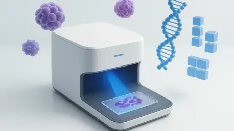

By identifying relevant microscopic patterns across large image patches, new AI models can process the immense complexity of whole slide images much faster than manual human observation. Modern computational pathology is undergoing a radical transformation as deep learning architectures move beyond simple cell counting toward the prediction of underlying genetic alterations. Traditionally, identifying specific mutations required expensive molecular testing, such as next-generation sequencing, which can take weeks to return results. However, recent breakthroughs in vision transformers allow researchers to extract subtle morphological features from routine H&E stained slides that are invisible to even the most experienced pathologists. These digital biomarkers correlate directly with driver mutations in lung and breast cancers, offering a glimpse into the tumor’s molecular profile within minutes. This shift represents a move toward integrated diagnostics where visual data serves as a surrogate for complex genetic mapping.
The Architecture of Predictive Pathology
The transition from traditional machine learning to advanced vision transformers has been pivotal in analyzing these high-resolution images. Unlike older models that struggled with the sheer scale of a whole slide image, these newer systems utilize attention mechanisms to focus on specific regions of interest that indicate malignancy. These models are trained on massive datasets where each digital slide is paired with its corresponding genomic sequence data. By learning these associations, the AI develops a refined understanding of how certain mutations, such as EGFR in lung cancer, manifest physically within the cellular architecture. This capability allows for a screening process where patients can be prioritized for specific targeted therapies almost immediately. Consequently, the reliance on tissue-heavy molecular assays is decreasing, as the digital scan provides a preliminary molecular blueprint that guides clinical decision-making without exhausting biopsy samples.
Integrating these AI-driven predictions into the standard laboratory workflow solves a significant logistical hurdle in oncology. Most community hospitals possess the equipment to digitize slides but lack the specialized facilities for comprehensive genomic profiling. By deploying these predictive models, these institutions can provide sophisticated diagnostic insights that were previously reserved for major academic medical centers. The software analyzes the spatial arrangement of cells and the shape of the nuclei to detect patterns that signify DNA mismatch repair deficiency. Furthermore, the objective nature of the AI reduces the inter-observer variability that often plagues manual grading systems. While a human pathologist might see a general pattern of inflammation, the algorithm identifies specific spatial signatures associated with immune checkpoint expression. This ensures that the diagnostic process is both faster and more consistent across different environments.
Strategic Integration for Precision Medicine
Clinical adoption of these tools is accelerating as validation studies demonstrate their accuracy in real-world settings. Researchers have successfully applied these models to identify therapeutic targets in rare cancers where molecular testing is not routinely performed. This democratization of precision medicine ensures that more patients receive personalized treatment plans based on their unique tumor biology. Moreover, the ability to predict mutations from standard slides facilitates the retrospective analysis of large clinical trial cohorts. By re-examining archival slides from 2026 to 2028, researchers are discovering new associations between morphological features and long-term treatment responses. This wealth of retrospective data is fueling the development of even more robust algorithms that can predict not just mutations, but also the likelihood of drug resistance. This proactive approach allows clinicians to adjust treatment strategies before a patient shows signs of relapse.
The implementation of AI-driven mutation prediction fundamentally altered the landscape of oncological care by streamlining the path from biopsy to treatment. Medical centers successfully integrated these digital platforms into their pathology departments, resulting in a measurable decrease in the time required to initiate targeted therapy. Stakeholders focused on establishing rigorous quality control standards to ensure the reliability of AI outputs across diverse patient populations. This transition necessitated a collaborative effort between bioinformaticians and pathologists to create a transparent framework for clinical use. Ultimately, the shift toward digital genomics provided a scalable solution to the growing demand for personalized medicine. Healthcare providers prioritized the acquisition of high-resolution scanning hardware and the training of staff to manage digital workflows. These steps ensured that the diagnostic infrastructure remained resilient and capable of supporting new therapeutic interventions.