
The biomechanics of rheumatic stenosis involve a fusion of the valve commissures that must be forcibly separated without damaging the surrounding soft tissue. This pathological condition typically arises as a late-stage complication of rheumatic fever, an inflammatory response triggered by streptococcal infections in childhood. Over several decades, this persistent inflammation causes the mitral valve leaflets—which control blood flow between the left atrium and the left ventricle—to become progressively thickened, scarred, and immobile. As the valve opening narrows, the heart is forced to exert significantly higher pressure to move blood through the body, eventually manifesting as debilitating symptoms such as chronic fatigue, severe shortness of breath, and congestive heart failure. Because this condition remains a pervasive public health crisis in many developing nations, identifying the most effective treatment path is essential for improving patient longevity and quality of life.
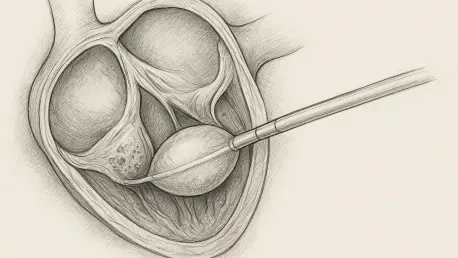
For many patients, the decision regarding intervention involves a choice between highly invasive open-heart surgery and the more modern, less invasive percutaneous transvenous mitral commissurotomy (PTMC). In the PTMC procedure, a cardiologist guides a specialized balloon-tipped catheter through the femoral vein into the heart chambers. Once the balloon is positioned within the stenotic mitral valve, it is rapidly inflated to crack the fused junctions and restore a more natural opening. While this approach offers the benefit of shorter hospital stays and a faster return to daily activities compared to a sternotomy, its success is not guaranteed. The structural integrity and the specific calcification patterns of the valve determine whether the balloon will cleanly separate the leaflets or cause traumatic damage to the heart’s delicate internal structures.
Limitations of Standard Diagnostic Tools
The Challenge: Subjectivity in the Wilkins Score
For the better part of four decades, the medical community has relied almost exclusively on the Wilkins score to determine if a patient is an appropriate candidate for a balloon-based valve opening. This echocardiographic grading system requires a sonographer to visually assess four distinct morphological features: the mobility of the leaflets, the degree of tissue thickening, the extent of calcification, and the health of the subvalvular apparatus, which includes the chordae tendineae. Each of these categories is assigned a score from one to four, and a cumulative score higher than eight generally suggests that the patient may face a higher risk of procedural failure or subsequent complications. However, as medical technology advances in 2026, the inherent limitations of this system have become more apparent to clinicians seeking higher precision.
The most significant drawback of the Wilkins score is its reliance on human interpretation, which introduces a high degree of subjectivity into the diagnostic process. Two different cardiologists may view the same ultrasound loop and arrive at different scores based on the angle of the probe or their individual clinical experience. Because ultrasound images can be grainy or obstructed by shadows, it is often difficult to distinguish between dense fibrous tissue and actual mineralized calcium. This lack of a truly quantitative, standardized measurement means that some patients who appear to be good candidates on paper actually possess hidden anatomical challenges that only become apparent once the procedure is already underway. Consequently, there is an urgent need for more objective imaging modalities that can provide a clearer mechanical picture of the valve.
Structural Obstacles: Mechanical Consequences of High Calcification
The presence of calcium within the mitral valve acts as a fundamental mechanical barrier that changes how the tissue responds to the outward force of a balloon. In a healthy valve suffering only from simple fusion, the balloon applies an even pressure that “pops” the leaflets apart at their natural seams. However, when calcium deposits are present, they act like rigid concrete reinforcements embedded within a flexible fabric. These mineralized zones do not stretch or yield; instead, they concentrate the mechanical stress into the adjacent non-calcified parts of the valve. If the calcium is situated near the edges of the leaflets, the force of the balloon is often diverted away from the fused junction and into the thin, vulnerable body of the leaflet itself.
This diversion of force frequently leads to the most dreaded complication of PTMC: the development of acute mitral regurgitation. When the valve leaflet tears instead of splitting at the commissure, blood begins to leak backward into the left atrium, creating a new and often more severe hemodynamic problem for the patient. Current ultrasound techniques struggle to map the exact density of these deposits, making it difficult to predict which valves will “snap” correctly and which will “shatter” under pressure. By failing to account for the specific volume and mass of the calcium, traditional scoring methods leave a significant margin for error. Understanding the spatial distribution of these hard deposits is therefore critical for moving beyond a trial-and-error approach to valve intervention.
Advancements in Quantitative Imaging
Technical Foundations: The Role of CT Mitral Valve Calcium Scoring
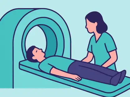
To address the lack of precision in traditional ultrasound, researchers have increasingly turned to electrocardiogram-gated computed tomography (CT) as a primary diagnostic aid. This technology allows for the capture of high-resolution images that are synchronized with the patient’s heart rhythm, effectively freezing the motion of the valve and providing a crystal-clear view of its structure. The most significant advantage of this modality is its ability to generate a Mitral Valve Calcium Score (MVCS) based on Hounsfield units. This is a standardized scale of radiodensity that allows computers to identify and quantify mineral deposits with a level of accuracy that far surpasses the human eye. Unlike the semi-quantitative nature of the Wilkins score, the MVCS provides a hard numerical value representing the total volume of calcium present.
By utilizing these objective measurements, clinicians can create a three-dimensional “mechanical map” of the mitral valve before the patient ever enters the operating room. This data allows the medical team to see not just where the calcium is, but how dense it is and how much total mass it occupies. In the current landscape of 2026, where precision medicine is the gold standard, this shift from visual estimation to digital quantification represents a major leap forward. The CT scanner can detect tiny micro-calcifications that ultrasound might miss, offering a more realistic assessment of how the valve will behave when subjected to the high-pressure inflation of a PTMC balloon. This level of detail is becoming indispensable for identifying patients who might otherwise have been subjected to a procedure with a low probability of success.
Evidence from the Field: Comparative Analysis of Clinical Research
A pivotal study conducted at the Rajaie Cardiovascular Institute recently provided concrete evidence regarding the predictive power of the MVCS in a clinical setting. The research team followed 30 patients with severe rheumatic mitral stenosis, each of whom underwent both a standard echocardiogram and a gated cardiac CT scan prior to their PTMC procedure. The study’s criteria for success were rigorous, requiring a significant increase in the valve’s surface area without the occurrence of a major leak. Upon analyzing the data, the researchers found a dramatic disparity between the two groups. Those who underwent a successful procedure had an average CT calcium score of just 0.76, whereas those in the failure group had a significantly higher average score of 16.08.
While the traditional Wilkins score was also higher in the failure group, it did not provide the same level of granular detail as the CT-derived data. The study highlighted that the presence of even moderate calcium density was a much stronger indicator of potential valve tearing than the mobility of the leaflets alone. Interestingly, the research also noted that in patients with a calcium score of zero, the success rate was nearly universal, suggesting that the MVCS could serve as a powerful “rule-in” tool for low-risk candidates. Although the small sample size meant that some statistical markers required further validation in larger cohorts, the numerical trends were so stark that they presented a compelling argument for the routine use of CT calcium scoring in the pre-operative planning phase.
Implications for Global Healthcare Delivery
Strategic Resource Management: Improving Patient Triage and Safety
The integration of CT calcium scoring into the clinical workflow offers a transformative opportunity for patient triage, particularly in regions where healthcare resources are stretched thin. In many developing nations, the ability to perform open-heart surgery is limited by the availability of specialized surgeons and intensive care facilities, making PTMC the primary treatment option. However, performing a balloon procedure on a high-risk patient with heavy calcification often results in failure or the need for emergency surgery—a scenario that can be catastrophic in a low-resource environment. By using the MVCS to identify these high-risk individuals early, medical teams can avoid the costs and risks associated with unsuccessful interventions and instead focus their efforts on medical management or referral to surgical centers.
Beyond the immediate financial benefits, the adoption of objective imaging significantly enhances patient safety by reducing the incidence of procedural complications. When a cardiologist knows the exact location and density of calcium deposits, they can make more informed decisions about the size of the balloon and the degree of pressure applied during inflation. This proactive approach minimizes the likelihood of causing a traumatic tear in the valve tissue, which often requires immediate and complex surgical correction. As CT technology becomes more accessible and cost-effective, the ability to extract predictive data from a standard scan provides a high-value diagnostic “add-on” that does not increase the patient’s radiation burden or financial strain, provided the scan is already being performed for other diagnostic reasons.
Precise Intervention: Future Protocols for Precision Valve Repair
Moving forward, the validation of CT calcium scoring is set to redefine the global standards for treating rheumatic heart disease. The ability to predict the fate of a heart valve procedure with such high accuracy allows for the development of personalized treatment plans that are tailored to the unique anatomy of each patient. As larger, multi-center trials continue through 2026 and into 2027, the medical community will likely establish definitive calcium score thresholds that will be written into international clinical guidelines. These thresholds will empower clinicians to provide clearer expectations to patients and their families, ensuring that the chosen path of treatment is based on hard data rather than professional intuition.
The researchers at the Rajaie Cardiovascular Institute demonstrated that while the traditional Wilkins score remains a foundational tool, it is no longer sufficient on its own in the era of high-definition imaging. The transition to a more objective model of assessment allowed cardiologists to anticipate mechanical failures before they occurred, shifting the focus from reactive complication management to proactive procedural planning. By the end of the study period, the medical team observed that incorporating the MVCS led to a more efficient use of the catheterization lab and a noticeable improvement in overall patient outcomes. This shift toward quantitative diagnostic protocols represented a significant milestone in the ongoing effort to eradicate the long-term morbidity associated with rheumatic heart disease.