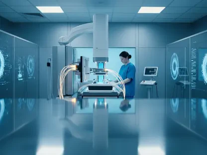

The intricate network of blood vessels and nerve fibers visible through a standard eye examination may hold the secret to identifying brittle bones years before a single fracture occurs. While most people visit their eye doctor to correct blurry vision or check for glaucoma, researchers are now proving that the back of the eye serves as a high-fidelity mirror for the entire body’s internal aging process. By leveraging the power of deep learning, medical science is beginning to translate the subtle map of the retina into a predictive tool for skeletal health, transforming a routine vision check into a comprehensive biological audit.
Could the Window to Your Soul Also Be a Window to Your Skeletal Health?
Osteoporosis has long been described by clinicians as a “silent thief” because it works in the shadows, stripping away bone density without causing any physical sensation until a sudden, painful break occurs. For many older adults, the first sign of the disease is a fractured hip or wrist resulting from a minor stumble, by which point the damage to the skeletal architecture is already severe. However, the tiny microvasculature in the retina shares many of the same biological pathways as the vascular systems supporting our bones, meaning that early signs of decay often appear in the eye first.
This connection offers a radical new perspective on preventative medicine. Instead of waiting for clinical symptoms or late-stage bone loss, doctors can now look toward the neural and vascular tissues at the back of the eye for whispers of future trouble. This specialized scan does not just look for eye disease; it looks for the physiological signatures of systemic aging. By identifying these markers early, healthcare providers can intervene with lifestyle changes or medical treatments long before the patient reaches a crisis point.
The transition from traditional eye care to systemic health monitoring is driven by the realization that the body does not age in isolated compartments. If the blood vessels in the eye show signs of accelerated wear and tear, it is highly probable that the skeletal and cardiovascular systems are following a similar downward trajectory. This holistic understanding of aging is the foundation of a new era in diagnostics where the eye becomes the ultimate diagnostic dashboard.
The Diagnostic Gap: The Hidden Crisis of Bone Health
Despite its prevalence, osteoporosis remains one of the most underdiagnosed conditions in modern healthcare, affecting nearly one in five people globally. This figure is frequently cited as a conservative estimate because the current diagnostic infrastructure is not designed for mass, routine screening. The “diagnostic gap” refers to the millions of individuals who are losing bone mass every day but fall through the cracks of the healthcare system because they do not meet the strict, often age-dependent, criteria for specialized testing.
The current gold standard for measuring bone density is the Dual-energy X-ray Absorptiometry (DEXA) scan. While highly effective at measuring mineral content, DEXA machines are expensive, require specialized technicians, and expose patients to a small amount of radiation. Consequently, these scans are typically reserved for patients who are already considered high-risk, such as post-menopausal women or those with a family history of fractures. This reactive approach means that a vast majority of the population remains unmonitored until it is too late to prevent the first injury.
To solve this crisis, there is a desperate need for a non-invasive, low-cost screening tool that can be deployed in a primary care or optometry setting. A tool that requires no radiation and utilizes existing imaging equipment could democratize bone health monitoring. By moving the initial screening process from a specialized radiology department to a local clinic, the medical community can finally address the massive backlog of undiagnosed cases and shift the focus from fracture management to proactive prevention.
Bridging the Gap: Ophthalmology and Orthopedics Through AI
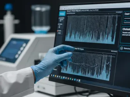
The technological bridge between these two seemingly unrelated fields is a deep-learning model known as RetiAGE. This artificial intelligence system analyzes retinal fundus images to calculate a person’s biological age—a metric that is often far more telling than chronological age. While your calendar age counts the years since your birth, your biological age measures the actual physiological condition of your tissues. If your retina appears ten years older than your birth certificate suggests, it indicates that your body’s internal systems are aging at an accelerated rate.
The retina is a unique anatomical site because it provides the only non-surgical view of the body’s microvasculature and neural tissue. Because the biological mechanisms that cause blood vessels to stiffen or nerves to degenerate in the eye are identical to the processes happening in the bone marrow and skeletal system, the “retinal age gap” serves as a proxy for the health of the entire body. The AI can detect patterns of thinning or vascular changes that are invisible to the human eye, translating these pixels into a risk score for osteoporosis.
This AI-driven approach was rigorously tested using the VGG16 convolutional neural network, which processed over 129,000 images to learn the subtle hallmarks of biological aging. By comparing these images against the health records of tens of thousands of participants, the researchers developed a high-confidence model capable of identifying systemic decay. This breakthrough effectively turns a simple photograph of the eye into a data-rich report on the state of a person’s skeletal durability.
Proving the Connection: Global Data and Future Outcomes
The validity of the RetiAGE model was confirmed through extensive analysis of two major international cohorts, starting with the Singapore PIONEER Study. In this group of nearly 2,000 older adults, researchers found that higher retinal age scores were directly correlated with lower bone mineral density in the hip and femur. For every standard deviation increase in the retinal age gap, the risk of experiencing a major osteoporotic fracture climbed by nearly 50%, providing clear evidence that the eye reflects the current state of the skeleton.
Even more impressive was the longitudinal data provided by the UK Biobank, which followed more than 43,000 participants over a 12-year period. At the start of the study, none of these individuals had a diagnosis of osteoporosis. However, those whose retinal scans showed signs of accelerated aging were 40% more likely to develop the disease over the following decade. This longitudinal proof demonstrates that the AI scan is not just a snapshot of current health, but a predictive tool capable of forecasting a diagnosis long before it is clinically detectable.
These findings highlight the potential for “opportunistic screening,” where data collected for one purpose—like an eye exam—is used to provide insights into other areas of health. By integrating these AI scores into existing medical workflows, doctors can gain a multi-system profile of a patient’s health. This research solidifies the retina’s role as a primary indicator of longevity, linking eye health to the risk of cardiovascular events, neurological decline, and now, the structural integrity of the skeletal system.
Frontier of Digital Health: Evidence From PLOS Digital Health
The research published in PLOS Digital Health suggests that adding AI-driven retinal scores to existing assessment tools, such as the Osteoporosis Self-assessment Tool, significantly sharpens diagnostic accuracy. By increasing the Concordance index and improving the reclassification of high-risk individuals, the RetiAGE model adds a layer of objective data that traditional medical histories and lifestyle questionnaires often miss. This is particularly valuable for patients who may not have obvious risk factors but possess a genetic or biological predisposition to rapid aging.
Furthermore, this study reinforces the idea that digital health tools can act as a powerful frontline filter for more intensive medical procedures. By using a quick, non-invasive eye photo to identify those at the highest risk, healthcare systems can prioritize who actually needs a DEXA scan. This ensures that expensive and specialized resources are used where they are most needed, increasing the overall efficiency of the healthcare system while reducing the burden on both patients and providers.
Medical experts believe this represents a paradigm shift in how we approach chronic disease. The ability to use a single image to assess multiple organ systems is the hallmark of the next generation of precision medicine. As the technology continues to mature, the integration of retinal aging scores could become a standard part of any annual physical, providing a continuous “health weather report” that tracks how well a person is aging over time.
Implementing Opportunistic Screening: The Path Forward
To bring this technology into the mainstream, healthcare systems must begin integrating AI analysis software into the retinal cameras already present in most optometry offices and primary care clinics. This integration would allow patients to receive a bone health risk assessment during their standard vision check, eliminating the need for additional appointments or travel to specialized imaging centers. It effectively turns every optometrist into a frontline guardian of skeletal health, catching the early warning signs of osteoporosis during the course of routine care.
Once a high RetiAGE score is detected, the patient can be fast-tracked for a formal DEXA scan to confirm their bone density levels and begin treatment if necessary. This workflow creates a seamless transition from screening to diagnosis, ensuring that those at high risk do not slip through the diagnostic gap. Additionally, because biological age is dynamic, tracking these scores annually could allow doctors to monitor the effectiveness of interventions like weight-bearing exercise, vitamin D supplementation, or pharmacological treatments.
The transition toward this model required a fundamental shift in how medical professionals approached preventative screenings and data sharing across specialties. Healthcare administrators recognized the necessity of establishing standardized protocols for AI integration to ensure that retinal data was interpreted consistently across diverse clinical settings. By prioritizing the calibration of these algorithms for different ethnic populations and camera types, the medical community successfully moved toward a more equitable and accurate diagnostic landscape. This evolution helped transform the standard eye exam into a vital component of holistic longevity management, ensuring that bone health was no longer a silent concern but a visible priority.