
Ivan Kairatov stands at the forefront of biopharmaceutical innovation, possessing an intricate understanding of how cutting-edge physics can be harnessed to solve the most pressing mysteries in cellular biology. With a career dedicated to research and development, he has spent years bridging the gap between engineering and medicine to visualize the invisible. In this conversation, we explore a transformative nanoscopy technique known as RO-iSCAT, which allows scientists to peer into the secret, dynamic lives of living cells in three dimensions without the use of harmful dyes.
How does rotating the angle of light illumination specifically strip away background noise to reveal 3D structures? What were the main engineering challenges in achieving a tenfold increase in the light signal while maintaining a live imaging environment over several days?
The beauty of rotating illumination lies in its ability to selectively isolate the signal coming from the cell while discarding the chaotic glare of the background. By sweeping the angle of light, we essentially perform a real-time subtraction of optical “clutter,” allowing the three-dimensional architecture of the cell to emerge with sudden, sharp clarity. The engineering feat here was significant because we had to boost a nearly undetectable bounce of light by tenfold without flooding the sample with energy. Maintaining this balance over several days required a incredibly “gentle” imaging protocol to ensure the light didn’t heat or stress the cells. This long-term stability is what finally allowed us to witness the continuous lifecycle of nanoscale structures rather than just taking a fleeting snapshot.
Traditional nanoscopy often relies on chemical dyes that can be toxic to living cells. How does removing these labels change the biological accuracy of what is observed, and what specific behaviors are now visible that were previously obscured by phototoxicity?
When you remove chemical dyes, you are essentially removing a mask that has historically skewed our understanding of cellular health. Labels often cause phototoxicity, where the light used to excite the dyes creates free radicals that damage or even kill the cell, leading to “unnatural” behaviors that scientists might mistake for biology. By using a label-free approach, we are seeing the cells in their most authentic state, free from the stress of chemical interference. This has revealed highly sensitive processes, such as the delicate retraction and reconnection of membrane protrusions, which would have likely collapsed under the harsh conditions of traditional fluorescent imaging. We are now seeing the “secret life” of cells exactly as it happens in the human body, with a level of biological accuracy that was previously impossible.
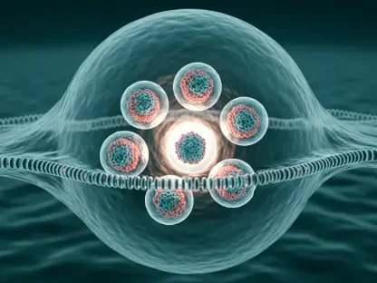
Cell extensions are now seen twisting and forming stable bridges rather than remaining static. Could you walk us through the mechanics of this dynamic motion and explain how these bridges facilitate the transfer of biochemical messages between neighboring cells?
The motion we captured is far more athletic and intentional than the scientific community previously assumed. These thin, thread-like extensions don’t just drift toward one another; they exhibit a highly dynamic twisting motion, almost like two vines winding together for support. Once they make contact, they fuse into a stable bridge that acts as a physical highway for the exchange of critical information. These structures allow cells to pass biochemical signals directly to their neighbors, ensuring that the entire tissue responds as a single, coordinated unit. It is a sophisticated communication network where the architecture itself is constantly being rebuilt and rerouted to meet the needs of the cellular population.
Pancreatic cancer cells and blood vessels form tight bridges with connective tissues to aid tumor growth. How can mapping these specific nanoscale pathways lead to more precise drug delivery, and what role do these bridges play in the way viruses migrate between cells?
Mapping these bridges gives us a literal “map of the enemy’s supply lines,” particularly in aggressive environments like pancreatic cancer. We have observed cancer cells forming multiple tight bridges with surrounding connective tissue and blood vessels, which helps the tumor reshape its environment to resist treatment or fuel its own growth. If we can identify the specific nanoscale pathways these bridges use, we can design drug delivery systems that “hijack” these routes to send therapies directly into the heart of the tumor. Furthermore, these bridges are suspected to be the primary corridors for viral migration, allowing viruses to hop from one cell to another while staying hidden from the immune system. Understanding the mechanics of these connections is the first step toward learning how to block them entirely.
What is your forecast for the future of nanoscale cellular imaging in disease treatment?
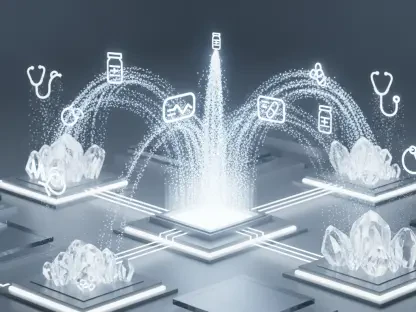
I believe we are entering an era where nanoscale imaging will move from the research lab into the heart of personalized medicine and drug development. In the coming years, the ability to monitor how a patient’s specific cells—such as their own human blood vessel cells or tumor cells—react to a therapy in real time will become a gold standard for efficacy. We will likely see the development of high-throughput screening tools that use this rotational light technique to test thousands of compounds, looking specifically for those that can disrupt the bridges formed by pathogens or cancers. Ultimately, our ability to visualize these 3D interactions within large cell populations will lead to a new generation of “smart” therapies that are precisely tuned to the structural vulnerabilities of a disease, saving more lives through pure, optical precision.