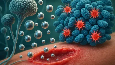

A critical challenge in modern healthcare involves overcoming the mobile genetic resistance determinants that allow MRSA to form resilient and treatment-resistant biofilms. As clinical settings navigate the complexities of 2026, the persistent threat of methicillin-resistant Staphylococcus aureus continues to undermine surgical outcomes and dermatological health globally. Standard antimicrobial therapies are frequently rendered obsolete by the rapid evolutionary pace of these bacterial colonies, which utilize complex genetic signaling to shield themselves from pharmaceutical intervention. This deadlock has catalyzed a shift toward “green” nanotechnology, an innovative field that merges biological efficiency with materials science. By utilizing fungal “biofactories” such as Fusarium solani, scientists have developed a method to produce silver nanoparticles that bypass the toxicity and environmental costs of traditional chemical synthesis. These biogenic particles do not merely act as passive antiseptics; they serve as dynamic therapeutic agents capable of dismantling bacterial defenses while simultaneously orchestrating the physiological processes required for rapid tissue regeneration in infected hosts.
Fungal Selection and the Biological Synthesis Pathway
The successful creation of these therapeutic nanoparticles began with the careful selection of a biological source capable of surviving in competitive microbial environments. Researchers focused their efforts on the rhizosphere—the biologically active soil layer surrounding plant roots—within agricultural zones in Egypt. From this organic-rich habitat, five distinct fungal isolates were retrieved and screened for their ability to convert silver ions into metallic nanoparticles. Among these candidates, the soil fungus Fusarium solani emerged as the most proficient producer, demonstrating a unique ability to synthesize particles with high antibacterial potency. This specific isolate was rigorously identified using internal transcribed spacer (ITS) ribosomal DNA sequencing, which confirmed a perfect genetic match with established databases. This biological foundation ensures that the production process remains consistent and reproducible, utilizing a fungus that is naturally adapted to producing high volumes of secondary metabolites in nutrient-rich soil conditions.
The synthesis itself is a sophisticated example of natural biochemistry, relying on the extracellular filtrate of the fungus rather than harsh industrial reducers like sodium borohydride. Detailed analysis of this filtrate revealed a complex mixture of compounds, including volatile organics, nitrogen heterocycles, and specific phenolic derivatives. A key player in this process is 2,3-dihydroxybenzoic acid, which contains a catechol moiety that facilitates the donation of electrons needed to reduce monovalent silver ions into zero-valent metallic silver. Simultaneously, long-chain fatty acids, including oleic acid, function as “capping agents.” These biological molecules form a protective shell around each nanoparticle, preventing aggregation and ensuring the stability of the colloidal suspension. This natural coating not only maintains the particles in a usable state but also enhances their biocompatibility when introduced into mammalian systems, avoiding the irritation often associated with chemically synthesized counterparts.
Physicochemical Characterization of Biosynthesized Silver
To validate the structural integrity of the newly formed nanomaterial, the research team employed a comprehensive suite of analytical tools to define the physical properties of the silver particles. The initial indicator of a successful synthesis was a clear transition in color from pale yellow to a deep, dark brown, a visual marker of surface plasmon resonance. This was further confirmed through ultraviolet-visible (UV-Vis) spectroscopy, which identified a sharp absorption peak at 410 nanometers, a signature characteristic of spherical silver nanoparticles. Furthermore, X-ray diffraction patterns revealed that the silver was arranged in a crystalline face-centered cubic lattice, proving that the biological method produces a high-quality metallic core. These characterization steps are essential for ensuring that the nanoparticles meet the strict manufacturing standards required for potential clinical applications in wound care and infection management.
Beyond the core structure, the size and electrostatic charge of the particles play a defining role in how they interact with biological tissues and bacterial cell walls. Using dynamic light scattering, researchers measured a Z-average hydrodynamic diameter of approximately 121.4 nanometers. However, high-resolution transmission electron microscopy showed that the actual metallic silver cores were much smaller, averaging only 19.58 nanometers. This discrepancy is attributed to the “biological crown” of fungal proteins and polysaccharides that surround the metal core, which increases the effective size in liquid environments. The particles also maintained a strong negative zeta potential of -42.7 millivolts. This significant negative charge is critical for long-term storage and clinical efficacy, as it provides the electrostatic repulsion necessary to keep the particles evenly dispersed and prevents them from settling or clumping into inactive masses during treatment application.
Mechanisms of Action Against Resistant Strains
The antibacterial efficacy of the Fusarium-derived nanoparticles was put to the test against ten clinical isolates of MRSA, all of which were molecularly confirmed to carry the mecA gene, the primary driver of resistance to methicillin and other beta-lactam antibiotics. In laboratory assays, the nanoparticles demonstrated impressive potency, with minimum inhibitory and bactericidal concentrations ranging from 0.1875 to 0.375 millimolar. These results indicate that the particles do not simply stunt the growth of the bacteria but effectively eliminate the population. This bactericidal capability is a significant advancement over many traditional antibiotics that have lost their edge against MRSA. The ability to neutralize these pathogens at such low concentrations suggests that biogenic silver could serve as a powerful alternative or supplement to current frontline treatments in hospitals dealing with high rates of multidrug resistance.
The specific mechanism of destruction was visualized through high-resolution microscopy, providing a clear picture of how the silver particles defeat MRSA. Upon contact, the nanoparticles appear to cause catastrophic structural failure in the bacterial cell envelope. The cell walls of treated bacteria were observed to become wavy and deformed, with the thick peptidoglycan layer completely detaching from the underlying plasma membrane. This physical disruption leads to the uncontrollable leakage of internal cellular contents, including vital proteins and DNA, resulting in rapid cell death. Confocal laser scanning microscopy further supported these findings, showing that a short two-hour exposure to the nanoparticles reduced the viability of MRSA populations from over 92 percent to less than 28 percent. This rapid onset of action is vital for preventing the bacteria from mounting a defensive response or further fortifying their protective biofilms.
Therapeutic Outcomes in Cutaneous Wound Recovery
Transitioning from laboratory testing to a living mammalian model, the researchers evaluated the performance of the silver nanoparticles in an excisional cutaneous wound model using rats. In this phase of the study, infected wounds were treated with a topical application of the biogenic particles to observe their impact on the healing timeline. The results were visually and statistically profound; by the 14th day of treatment, the nanoparticle-treated group achieved a 95.33 percent wound closure rate. This stands in stark contrast to the untreated infected group, which showed a closure rate of only 12.33 percent, leaving the wounds open, inflamed, and prone to further complications. This level of acceleration in tissue repair highlights the particles’ potential to transform the management of chronic, infected ulcers that otherwise might take months to heal or lead to systemic illness.
The success of the treatment was also reflected in the microbiological data collected from the skin tissue of the animal subjects. In the untreated rats, the bacterial burden remained extremely high at 9.90 log CFU/g, indicating a persistent and aggressive infection. However, in the rats that received the silver nanoparticle treatment, the bacterial load was slashed to just 1.10 log CFU/g. This near-total clearance of the MRSA infection allowed the body’s natural regenerative cycles to proceed without the persistent interference of pathogenic toxins and inflammatory signals. By removing the primary biological barrier to healing, the nanoparticles enabled the wounds to move quickly through the inflammatory phase and into the proliferative phase of repair. This dual capability—eradicating resistant bacteria while clearing the path for tissue growth—positions biogenic silver as a comprehensive solution for complex wound management.
Modulation of Chronic Inflammatory Environments
One of the most significant insights gained from this research was the ability of the silver nanoparticles to actively modulate the host’s immune response. In chronic or infected wounds, the body often gets trapped in a cycle of overactive inflammation, characterized by high levels of pro-inflammatory cytokines like interleukin-6 (IL-6) and tumor necrosis factor-alpha (TNF-alpha). This persistent inflammation causes further tissue damage and prevents the formation of healthy new skin. The study revealed that application of the Fusarium-derived nanoparticles significantly reduced the levels of these inflammatory markers, as well as the expression of cyclooxygenase-2 (COX-2). By dampening these signals, the nanoparticles helped “reset” the wound environment, shifting it from a state of destructive defense to one of active reconstruction and tissue synthesis.
Histological analysis of the treated skin tissue confirmed that this reduction in inflammation was accompanied by superior structural regeneration. The wounds treated with silver nanoparticles showed more organized dermal layers, better epidermal resurfacing, and significantly improved vascularization compared to the controls. The formation of new blood vessels is a cornerstone of effective healing, as it ensures the delivery of oxygen and nutrients to the site of repair. In the treated group, the tissue reconstruction was so effective that the skin layers began to resemble healthy, uninjured tissue within the two-week study period. This immunomodulatory effect suggests that the nanoparticles do more than just clean the wound; they interact with the biological signaling pathways of the host to promote a more efficient and less scarred recovery process.
Integrating Green Nanotechnology into Modern Medical Practice
The comprehensive evaluation of Fusarium solani silver nanoparticles demonstrated that these biogenic materials provided a viable and sustainable pathway for treating resistant infections and accelerating wound recovery. This study highlighted how the natural enzymatic processes of fungi were harnessed to create highly stable, biocompatible agents that outperformed traditional expectations in both antibacterial and regenerative roles. It was established that the particles significantly reduced pro-inflammatory markers and bacterial loads in living tissue, which facilitated a faster transition to the repair phase of healing. These findings suggested that the integration of biological synthesis into the pharmaceutical supply chain could alleviate the environmental burden of chemical manufacturing while offering a potent new tool for the 2026 to 2028 healthcare landscape.
Moving forward, the industry must prioritize several key steps to translate these laboratory successes into widespread clinical use. Standardized toxicity profiles and detailed biodistribution studies remained essential to ensure that topically applied silver does not accumulate in internal organs over long treatment durations. Future research efforts were directed toward comparing these green nanoparticles directly with frontline antibiotics like vancomycin to quantify their relative advantages in clinical settings. Furthermore, healthcare systems were encouraged to explore the large-scale cultivation of fungal biofactories as a cost-effective means of producing advanced therapeutics. By addressing these technical and safety hurdles, the medical community moved closer to a future where eco-friendly nanotechnology serves as a cornerstone of infection control and personalized wound care.