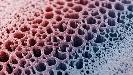

The historical quest to engineer fully functional human organs has long been obstructed by the immense complexity of replicating the microscopic vascular networks that sustain living tissue. While modern science has mastered the art of printing large-scale arterial structures, the delicate web of capillaries—those microscopic conduits responsible for the essential exchange of oxygen and nutrients—has remained an elusive target for fabrication. These tiny vessels, often narrower than a single human hair, are the biological workhorses that prevent tissue death by ensuring that every cell within a three-dimensional mass has access to the circulatory system. Without a viable method to create these intricate pathways, the production of transplantable, lab-grown organs remains limited to thin sheets of tissue that cannot survive in a complex, multi-layered state. Recent advancements in hybrid bioprinting and artificial intelligence are now offering a pathway toward solving this critical bottleneck in medical science.
Barriers to Progress in Microvascular Engineering
Understanding the limitations of current technology is essential for appreciating the recent shift toward hybrid bioprinting systems. Traditional extrusion-based methods, which function by pushing a biological ink through a nozzle, have been the industry standard due to their speed and ability to handle high concentrations of cells. However, these systems are fundamentally limited by the physical size of the printer head; a nozzle small enough to print a capillary would frequently clog or subject delicate living cells to lethal amounts of shear stress. Most extrusion printers are unable to produce features smaller than several hundred microns, which is far too large for the sub-ten-micron scale required for true capillary function. This spatial gap has historically meant that engineered tissues lacked the dense irrigation required to support metabolic activity in thick, three-dimensional constructs, leading to core necrosis in larger samples.
In addition to physical constraints, the manufacturing process for vascularized tissue has suffered from a lack of scalability and precision in high-resolution printing. Techniques such as light-based stereolithography can achieve the necessary detail for microscopic vessels, but they are often prohibitively slow when tasked with building an entire organ. These methods also struggle to integrate the diverse range of materials needed to mimic the complex mechanical properties of natural tissue. Furthermore, the traditional reliance on manual trial-and-error to calibrate these systems has led to significant inconsistencies in output. Scientists have spent years attempting to balance the viscosity of bio-inks with printing speed and light intensity, only to find that minor environmental changes can ruin an entire production run. This inefficiency has highlighted the urgent need for a more intelligent, adaptable, and integrated manufacturing approach.
Precision and Power: The Hybrid Multi-Modality Model
The development of a dual-modality printing strategy represents a significant technological leap by merging aerosol jet printing (AJP) with traditional extrusion techniques. In this new workflow, AJP is utilized to deposit sacrificial materials with extreme precision, creating temporary scaffolds that define the paths of the smallest capillaries. This method utilizes an ultrasonic atomizer to create a fine mist of droplets, allowing for the deposition of lines that are thinner than ten microns without the risks of clogging associated with standard needles. These sacrificial patterns act as a high-fidelity blueprint for the microvasculature, providing a temporary structure that can be surrounded by more robust biological materials. Once the primary tissue structure is solidified, these temporary templates are dissolved or washed away, leaving behind a perfectly formed network of hollow channels that mimic the natural architecture of a human circulatory system.
While the aerosol jet printer manages the intricate details of the capillary bed, the extrusion printer provides the necessary volume and structural support for the surrounding tissue. This larger-scale system deposits the bulk of the cellular matrix, ensuring that the finished product has the mechanical integrity required for handling and potential surgical implantation. By combining these two modalities into a single integrated system, researchers can now create hierarchical vascular networks that transition seamlessly from large transport vessels to microscopic exchange sites. This hierarchical approach is vital because it ensures that blood can flow from a central artery into the smallest branches of the tissue without experiencing dangerous pressure drops or leakage. The result is a synthetic tissue construct that possesses both the durability of a macroscopic organ and the delicate functionality of a living biological system.
Machine Learning and the Automated Calibration Process
The successful coordination of multiple printing heads and diverse materials is made possible through the integration of a sophisticated artificial intelligence algorithm. Researchers have successfully implemented constrained Bayesian optimization to automate the complex calibration processes that previously required months of manual labor. This AI-driven approach creates a mathematical model of the printing process, allowing the system to learn from each successive attempt and make real-time adjustments to parameters such as pressure, speed, and temperature. Instead of relying on a human operator to guess the ideal settings for a specific bio-ink, the algorithm identifies the optimal configuration with minimal waste. This ensures that the intricate capillary patterns printed by the aerosol jet are consistent every time, regardless of the variations in material batches or environmental conditions.
The shift from manual calibration to autonomous optimization has significantly enhanced the scalability of bioprinting operations. By utilizing machine learning, the system can adapt to new biological requirements on the fly, allowing for the rapid prototyping of different tissue types and vessel geometries. This capability is particularly important when working with patient-specific cells, which can vary in their behavior and physical properties. The AI manages the complex interplay between the sacrificial inks and the structural matrices, ensuring that the interface between the two is seamless and that the final vascular channels are free of defects. This level of computational control reduces the error rate in tissue fabrication, moving the technology closer to the rigorous standards required for clinical applications. The integration of AI has effectively transformed bioprinting from an artisanal craft into a precise and repeatable engineering discipline.
Biological Validation and Future Clinical Implications
A vascular network must be more than just a series of hollow tubes; it must be biologically active and capable of regulating fluid flow. To validate the effectiveness of their hybrid system, researchers seeded the printed micro-channels with human endothelial cells, which are the primary building blocks of natural blood vessel linings. These cells successfully adhered to the walls of the channels, forming a continuous and healthy monolayer that replicated the internal surface of a real capillary. Most importantly, tests revealed that these optimized vessels exhibited a significant reduction in permeability compared to those produced by traditional methods. This barrier function is critical for preventing the leakage of fluids into the surrounding tissue, ensuring that the engineered vasculature can effectively transport oxygen and nutrients while maintaining the pressure needed for proper circulation within a living body.
The successful creation of functional capillary networks in the lab provided a robust foundation for a variety of transformative medical applications. These advanced vascular models were used to develop high-fidelity drug screening platforms that offered a more accurate representation of human physiological responses than traditional animal models. By observing how new pharmaceuticals interacted with functional human capillaries, scientists were able to identify potential side effects and efficacy issues much earlier in the development cycle. Looking forward, this technology was instrumental in the creation of personalized organ-on-a-chip devices, which allowed for the study of specific vascular diseases in a controlled environment. The ability to print intricate, life-sustaining networks moved the industry significantly closer to the ultimate goal of producing entire, transplantable human organs that could survive and thrive within the human body.