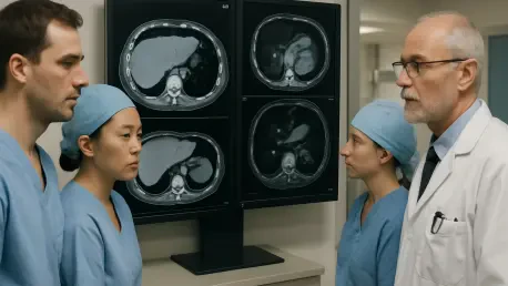

The clinical landscape of hepatobiliary surgery has undergone a profound transformation as medical teams increasingly rely on high-resolution imaging to evaluate donor organs before any surgical intervention begins. For decades, the decision to accept or reject a donor liver rested on subjective visual assessments and invasive biopsies that, while useful, often failed to capture the full physiological profile of the tissue. This high-stakes environment demands absolute precision, as the margin for error in matching a graft to a recipient is incredibly narrow. Recent research led by specialists in Geneva emphasizes that pre-retrieval imaging is no longer an optional diagnostic but a primary clinical strategy that directly influences patient survival. By utilizing non-invasive modalities such as Computed Tomography and Magnetic Resonance Imaging early in the process, transplant teams can identify potential complications long before the donor enters the operating room, ensuring that every available organ is used to its maximum potential without compromising safety.
Quantifying Hepatic Steatosis with Advanced Modalities
Hepatic steatosis, characterized by an overabundance of fat within the liver cells, remains one of the most significant barriers to successful transplantation in the current medical era. When an organ with high fat content is transplanted, it faces a substantially higher risk of primary non-function or delayed graft function, which can be fatal for the recipient. While ultrasound has traditionally served as the initial screening tool due to its portability and low cost, it often lacks the necessary sensitivity to detect mild or moderate steatosis, particularly in donors with a high body mass index. Consequently, transplant centers have moved toward more objective measurements provided by Computed Tomography. By analyzing Hounsfield units to determine the liver-to-spleen ratio, clinicians can now quantify fat levels with a degree of standardization that was previously unattainable. This shift toward quantitative metrics allows for a more rigorous selection process, minimizing the guesswork associated with visual inspections.
The pursuit of even greater accuracy has led to the widespread adoption of Magnetic Resonance Imaging, specifically utilizing the Proton Density Fat Fraction technique. This method is now recognized as the gold standard for non-invasive fat quantification, as it provides measurements that correlate almost perfectly with histological findings from traditional biopsies. For living donor programs, where the safety of the healthy donor is just as critical as the outcome for the recipient, the precision of MRI is indispensable. It allows surgeons to exclude donors with high fat levels early in the evaluation phase, preventing unnecessary surgical risks. Furthermore, the ability to map the distribution of fat across the entire organ, rather than relying on a small biopsy sample that may not be representative, ensures a comprehensive understanding of the graft’s health. As these imaging technologies become more accessible, they are setting a new benchmark for donor evaluation that prioritizes objective data over clinical intuition.
Mapping Internal Architecture for Surgical Success
Navigating the intricate vascular and biliary anatomy of the liver is a primary challenge for any transplant surgeon, as anatomical variations are common and can lead to catastrophic intraoperative errors if not identified early. Computed Tomography Angiography has emerged as an essential tool for creating a detailed three-dimensional roadmap of the donor’s arterial and venous systems. By identifying anomalies such as replaced hepatic arteries or unconventional venous drainage patterns, the surgical team can plan the implantation phase with meticulous detail. This level of preparation reduces the “cold ischemia time,” which is the duration the organ remains outside the body, thereby improving the chances of immediate graft function. When the surgical team knows exactly what to expect before making the first incision, the procedural flow becomes significantly more predictable, and the risk of technical failure during the delicate process of vascular reconstruction is drastically minimized.
In addition to vascular mapping, visualizing the biliary tree through Magnetic Resonance Cholangiopancreatography is vital for preventing postoperative complications like bile leaks or strictures. These issues are among the most frequent causes of long-term morbidity in transplant recipients and often require multiple follow-up procedures to correct. Beyond identifying ductal variations, imaging plays a critical role in liver volumetry, which is the precise measurement of the organ’s size and weight. Using advanced software to calculate graft volume ensures that the recipient receives a sufficient amount of functional liver mass to avoid the dreaded “small-for-size syndrome.” This condition occurs when the transplanted liver is too small to meet the metabolic demands of the recipient, leading to rapid organ failure. By accurately matching the graft size to the recipient’s needs, and ensuring the living donor retains enough liver for a safe recovery, imaging provides a double layer of protection.
Integrating Radiomics and Machine Perfusion Strategies
The current evolution of transplant medicine is defined by the integration of imaging data into a dynamic decision-support framework that connects donor quality with specific preservation techniques. Rather than viewing a liver graft in isolation, modern protocols now link pre-retrieval imaging findings to the use of machine perfusion technologies. For instance, if a CT scan identifies a “marginal” liver with moderate fat content, the transplant team might choose to use normothermic machine perfusion to “test” the organ’s function in a controlled environment before implantation. This synergy between diagnostic imaging and advanced preservation methods is expanding the donor pool by allowing surgeons to safely utilize organs that would have been discarded in the past. This approach transforms a static assessment into a proactive management strategy, where imaging serves as the foundational data point that dictates how the organ will be handled from the moment of retrieval to the final stitch.
Artificial intelligence and the emerging field of radiomics are further pushing the boundaries of what can be learned from pre-retrieval scans by extracting data that is invisible to the human eye. These algorithms can analyze thousands of pixels to identify subtle patterns in tissue texture and vascular flow that correlate with specific clinical outcomes. By training these models on large datasets of past transplants, researchers have developed predictive tools that can estimate the likelihood of graft success with remarkable precision. This data-driven approach allows for a more nuanced evaluation of “extended criteria” donors, who might be older or have secondary health issues but still possess viable organs. As these AI-driven insights become a standard part of the transplant workflow, the industry is moving toward a future where every organ is assessed through a lens of total transparency, ensuring that no viable graft goes to waste while maintaining the highest possible standards for recipient safety.
Advancing Outcomes through Standardized Clinical Protocols
To fully realize the benefits of pre-retrieval imaging, the global transplant community has prioritized the standardization of imaging protocols across different medical regions. Disparities in how images are captured or interpreted can lead to inconsistencies in organ allocation, which is why the establishment of universal guidelines for Hounsfield unit measurements and fat fraction reporting is so essential. By creating a common language for organ quality, transplant centers can facilitate smoother inter-hospital transfers and improve the transparency of the donor registry system. This collaborative effort ensures that a liver retrieved in one city can be confidently accepted by a team in another, based on a reliable and standardized digital profile. The focus is shifting from simply having the technology to ensuring it is applied consistently, which is the key to reducing waitlist mortality and optimizing the use of every donated organ in the modern healthcare infrastructure.
The implementation of these advanced imaging strategies successfully bridged the gap between organ scarcity and surgical demand by providing a clearer understanding of graft viability. Medical institutions that invested in high-resolution pre-retrieval diagnostics reported a significant decrease in postoperative complications and a notable increase in the successful use of complex donor grafts. The transition toward a data-centric model ensured that surgeons were no longer forced to rely on limited information during critical moments. By the conclusion of the most recent clinical trials, the evidence supported a shift toward mandatory imaging for all deceased and living donors, marking a new era in hepatobiliary medicine. These advancements provided the necessary tools to navigate the complexities of organ transplantation with a level of confidence that was previously unimaginable, ultimately saving more lives through the power of precise visualization and rigorous analytical standards.


