
Determining the survival trajectory of a gastric cancer patient often relies on a microscopic examination of the tumor’s architectural spread, yet this conventional approach frequently misses the vital physiological narrative of the host. Researchers at the State University of Campinas have addressed this gap by developing a sophisticated imaging biomarker known as Visceral Muscle Density (VMD), which provides a comprehensive view of a patient’s metabolic health. By repurposing routine computed tomography (CT) scans, this approach allows physicians to move beyond the limitations of traditional staging and enter a realm of more holistic prognostic assessment. The integration of such biomarkers into standard clinical workflows represents a profound shift in how the medical community views cancer, transforming standard diagnostic images into rich datasets that reflect the interplay between systemic inflammation, nutrition, and disease. This methodology offers a personalized prediction, ensuring that clinical decisions are informed by unique biological resilience.
Deciphering the VMD Marker: A Window Into Metabolic Health
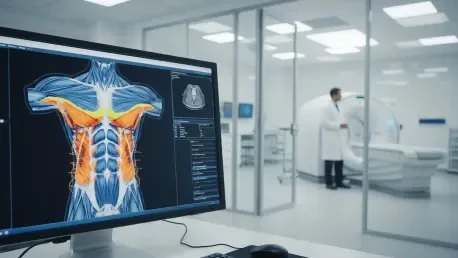
The Visceral Muscle Density marker is derived from the radiodensity of specific tissues, a measurement that reflects how effectively tissues like skeletal muscle and visceral fat attenuate X-rays during a CT scan. In clinical terms, radiodensity is expressed in Hounsfield Units, where higher values indicate denser tissue and lower values suggest the presence of infiltrates like fat or changes in tissue quality. High radiodensity in visceral fat is often indicative of systemic inflammation, a condition that can exacerbate tumor growth and weaken the body’s natural defenses against gastric cancer. Conversely, low radiodensity in skeletal muscle, often referred to as myosteatosis, signals a reduction in muscle quality and is a precursor to cachexia or muscle wasting. By combining these two distinct measurements into a unified indicator, clinicians can visualize an integrated phenotype that reveals the metabolic resilience of the patient, providing far more context than a biopsy could on its own.
This focus on host physiology recognizes that cancer does not exist in a vacuum but is instead deeply influenced by the patient’s internal environment and nutritional status. Traditional imaging has long been utilized to measure tumor dimensions, yet the wealth of data regarding the surrounding tissues has remained largely untapped until this recent push for advanced body composition analysis. The VMD marker acts as a functional proxy for the patient’s overall vigor, allowing oncologists to identify those who may be physically frail despite appearing otherwise healthy. Such insights are crucial because patients with poor metabolic profiles often suffer from increased toxicity during chemotherapy or higher rates of surgical complications. By shifting the prognostic lens from the tumor’s aggression to the host’s ability to withstand that aggression, the medical field is now better equipped to manage the complexities of gastric cancer. This approach ensures the biological reality of the patient is given equal weight.
Enhancing Data Reliability: The Intersection of AI and Radiomics
One of the persistent challenges in utilizing quantitative imaging data across multiple medical centers is the inherent variation between different CT scanner manufacturers and software configurations. These technical discrepancies can lead to inconsistent radiodensity readings, potentially compromising the accuracy of biomarkers like Visceral Muscle Density when applied in a large-scale clinical setting. To solve this problem, the research team developed a novel mathematical approach that utilizes the difference between fat and muscle density rather than relying on absolute values. By calculating this internal ratio, the researchers were able to cancel out the technical noise caused by different imaging hardware, ensuring that the VMD marker remains a robust and reliable tool regardless of where the scan was performed. This calibration technique is essential for the widespread adoption of the marker, as it allows for the seamless exchange of prognostic data between hospitals without needing specialized equipment.
Artificial intelligence has been the primary engine driving these advancements, enabling the analysis of vast datasets that would be impossible to process through manual observation alone. Machine learning algorithms were trained on thousands of clinical records and CT images to identify the exact tissue density patterns that correlate most strongly with patient survival and treatment response. These AI models can detect subtle nuances in tissue texture and density that the human eye might overlook, providing a level of precision that is necessary for accurate risk stratification. This synergy between radiomics and AI allows for the creation of predictive models that are continuously refined as more patient data becomes available, ensuring the VMD marker evolves with the latest clinical findings. Furthermore, this automated processing speeds up the diagnostic timeline, allowing clinicians to receive detailed reports within minutes. This integration not only enhances accuracy but also standardizes how clinicians interpret data.
Clinical Outcomes: Stratifying Risks and Personalizing Care
The impact of implementing the VMD marker in a clinical setting is perhaps most evident in the stark differences in survival rates observed among high-risk and low-risk patient groups. Clinical data indicates that patients identified as high-risk by the VMD marker experienced a median survival of approximately 13.8 months, a figure that pales in comparison to the 58.5 months observed in the favorable group. This nearly four-year gap in survival underscores the fact that body composition and metabolic health are foundational pillars of a patient’s overall prognosis, often carrying more weight than the tumor stage itself. Such a significant disparity highlights the danger of relying solely on tumor-centric metrics, which can lead to an overly optimistic or pessimistic outlook depending on the patient’s physical state. By utilizing the VMD marker, doctors can identify high-risk individuals early in the treatment process, allowing for more intensive monitoring and aggressive supportive care.
Beyond simple survival statistics, the VMD marker facilitates a more refined strategy known as therapeutic stratification, which involves tailoring the intensity of treatment to the patient’s physiological capacity. For instance, a patient with a poor VMD profile may be at higher risk for chemotherapy-induced toxicity, necessitating a modified dosage or the prioritization of supportive therapies to improve muscle density before starting aggressive regimens. Conversely, a patient with a strong metabolic profile might be a candidate for more intensive treatments that offer a better chance of long-term remission but require significant physical stamina. This level of customization ensures that the treatment plan is not just based on the type of cancer, but on the person who has the disease. Integrating nutritional interventions and physical rehabilitation into the oncology plan becomes a data-driven necessity, as clinicians now have the evidence to show how improving body composition can directly translate into better outcomes.
Advancing Precision Medicine: Implementation and Future Research
While the initial success of the Visceral Muscle Density marker in gastric cancer is promising, the journey toward global standardization and broader clinical application is just beginning. Ongoing research aims to validate this imaging biomarker across a wider range of ethnic populations and diverse geographic regions to ensure its effectiveness remains consistent regardless of genetic backgrounds. There is also a significant effort to investigate whether the VMD marker can be applied to other types of solid tumors, such as pancreatic or colorectal cancers, where metabolic health also plays a critical role in patient outcomes. By establishing a universal framework for measuring tissue density, the medical community can create a more cohesive approach to cancer care that prioritizes the host’s physiological state across various oncological disciplines. Furthermore, the cost-effective nature of this marker, which utilizes existing CT infrastructure, makes it an attractive option for healthcare systems in many developing nations.
The introduction of the VMD marker provided a significant breakthrough by offering a non-invasive and highly accessible method for predicting gastric cancer outcomes through standard clinical data. Clinicians successfully utilized these findings to implement targeted nutritional and exercise programs, aimed at improving tissue density and reducing systemic inflammation before patients underwent major surgeries. These proactive measures were complemented by the development of automated software modules that integrated seamlessly into existing hospital radiology platforms, ensuring that the VMD score was available to every member of the oncology team. Moving forward, the focus shifted toward longitudinal studies that monitored how changes in a patient’s VMD profile over the course of treatment served as a real-time indicator of therapy effectiveness. This dynamic approach allowed for the adjustment of treatment plans in response to the patient’s evolving health status. Ultimately, this innovation redefined the prognostic standard.


