
The election of Simon R. Cherry to the Royal Society marks a monumental milestone in the field of medical physics, distinguishing him as one of the few global leaders whose contributions have fundamentally altered the landscape of diagnostic imaging and clinical research. As a Distinguished Professor Emeritus at the University of California, Davis, Cherry joins an elite group of only ninety scientists worldwide selected for this honor in the current cycle, reflecting his status as a visionary in the integration of engineering and biology. His inclusion in the world’s oldest scientific academy is not merely a recognition of personal achievement but a testament to four decades of relentless pursuit in making the invisible visible through technological innovation. By pushing the boundaries of what was once considered scientifically impossible, he has provided the medical community with tools that offer unprecedented clarity into the human body’s inner workings, thereby setting a high standard for future researchers.
Diagnostic Precision: Advancing Care Through Molecular Imaging
Molecular imaging serves as the cornerstone of modern diagnostic medicine, allowing clinicians to observe biological processes in real time without resorting to invasive surgical procedures. Dr. Cherry’s work in this domain centered on the belief that biological events at the cellular level could be quantified and visualized through sophisticated instrumentation. Unlike traditional anatomical scans that show structure, molecular imaging provides a functional map of biochemical activity, offering a deeper understanding of how diseases like cancer and neurological disorders manifest and progress. This shift from structural to functional observation necessitated a complete rethinking of sensor technology and data reconstruction algorithms. By focusing on the molecular pathways, his research allowed for earlier detection of pathologies, often before physical symptoms or structural changes became apparent. This proactive approach to medicine has redefined the parameters of patient care in various clinical settings today.
In the effort to refine these diagnostic tools, Dr. Cherry addressed significant technical hurdles regarding the resolution of Positron Emission Tomography scans by developing the microPET scanner. This breakthrough utilized novel detector configurations to achieve high-resolution images previously thought unattainable, particularly for small-scale biological studies. This innovation was transformative for the pharmaceutical industry and academic research, as it allowed for the precise study of disease models with high fidelity. The ability to track drug interactions and disease progression with such accuracy accelerated the development of new therapeutics and streamlined the transition from laboratory discovery to clinical application. Today, the principles established by the microPET system remain foundational to the design of modern PET detectors, proving that engineering excellence can bridge the gap between abstract physics and practical medical utility. This legacy of precision continues to influence how the global scientific community approaches the complexities of internal biological systems.
Clinical Evolution: From Hybrid Systems to Total-Body Imaging
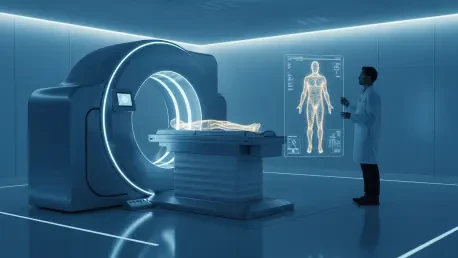
Building upon his success in high-resolution detectors, Dr. Cherry pioneered the integration of PET and Magnetic Resonance Imaging into a single, simultaneous system, despite widespread skepticism regarding the feasibility of such a fusion. By successfully operating radiation detectors within intense magnetic fields, he created hybrid scanners that provide a synchronized view of physiological processes and anatomical structures. This work culminated in the development of the EXPLORER, the world’s first total-body PET scanner, which can capture a head-to-toe image in a single acquisition. This device offers a sensitivity forty times greater than conventional scanners, allowing for significantly lower radiation doses and faster scan times for patients. To date, nearly one million patients have benefited from this technology, which provides a holistic view of systemic interactions across the entire body. The global reach of these systems confirms that integrated imaging is now an essential component of modern high-acuity healthcare, improving efficiency and safety simultaneously.
Looking toward future developments, researchers focused on the creation of ultrafast imaging detectors designed to reach picosecond levels of timing resolution. This effort aimed to enhance the signal-to-noise ratio in PET scans, allowing for the detection of even smaller metabolic anomalies with greater precision than ever before. The scientific community recognized that the success of these innovations was built upon a dedicated interdisciplinary philosophy that combined physics, biology, and engineering into a single cohesive strategy. Professionals in the field prioritized the practical application of these high-resolution tools to ensure they served as accessible solutions for clinicians worldwide. His election to the Royal Society solidified a legacy of transformation, as his career proved that the most effective way to advance human health involved a commitment to technological excellence and collaborative discovery. This groundwork established a clear path for future medical breakthroughs, ensuring that the next generation of scientists possessed the tools necessary to solve complex health challenges.