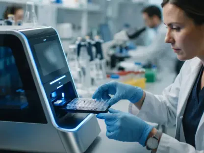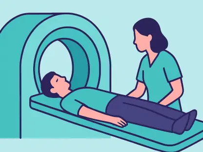

The rapid evolution of diagnostic precision across Western Australia has effectively turned the region into a global epicenter for medical innovation and advanced molecular imaging research. By integrating state-of-the-art cyclotron facilities with clinical practice, the state has managed to bridge the gap between experimental laboratory findings and real-world patient applications. This shift is not merely about purchasing expensive equipment but involves a coordinated ecosystem where researchers, clinicians, and government bodies align to solve complex diagnostic puzzles. The expansion of the Perth Health Precinct and the continued investment in the Harry Perkins Institute have solidified a foundation that supports high-resolution imaging techniques previously unavailable in the Southern Hemisphere. As the global demand for personalized medicine increases, Western Australia is utilizing its unique geographic and academic position to refine the accuracy of disease detection, ensuring that treatments are more targeted and effective for a diverse population.
Advancements in Radiopharmaceutical Production and Distribution
The establishment of sophisticated cyclotron facilities in Perth has revolutionized the local production of short-lived isotopes, which are essential for high-fidelity molecular imaging procedures. Previously, the logistical challenges of importing certain tracers meant that many diagnostic protocols were restricted by the half-life of the materials involved. Today, the production of Gallium-68 and Fluorine-18 occurs on-site, allowing for a seamless transition from synthesis to patient injection within the same medical complex. This localized capability ensures that clinicians have access to a reliable supply of radiopharmaceuticals, which in turn facilitates more frequent and precise PET/CT scans. Furthermore, the development of specialized cold kits for labeling molecules has simplified the preparation process, enabling smaller regional clinics to benefit from the high-end research conducted in the metropolitan core. This distributed network of expertise has significantly reduced the wait times for critical cancer staging and neurological assessments.
Beyond the basic production of isotopes, the focus has shifted toward the creation of novel ligands that can target specific biological markers with unprecedented sensitivity. Researchers at the University of Western Australia are currently pioneering the use of targeted alpha therapy, which combines diagnostic imaging with direct cellular treatment. This dual-purpose approach, often referred to as theranostics, relies on the ability of molecular imaging to identify exactly where a tumor is located and whether it expresses the specific receptors required for treatment. By utilizing Lutetium-177 and other emerging isotopes, Western Australian specialists can visualize the distribution of a drug throughout a patient body before the full therapeutic dose is administered. This methodology significantly lowers the risk of systemic toxicity and ensures that only the intended tissues are affected by radiation. The integration of these advanced chemical processes into the clinical workflow demonstrates a profound commitment to moving beyond traditional medical interventions.
Strategic Integration of Imaging Technology and Clinical Data
The deployment of total-body PET scanners, specifically the uEXPLORER system, provided a comprehensive view of the human body in a single frame, significantly outperforming traditional modular scanners. To manage the massive datasets generated by these high-resolution systems, Western Australian institutions integrated sophisticated artificial intelligence algorithms for image reconstruction and interpretation. These AI systems filtered out background noise and highlighted subtle metabolic changes that might be missed by the human eye during standard reviews. By training these models on thousands of localized cases, the software became attuned to specific demographic variations and common disease patterns found within the regional population. Machine learning tools also allowed for the prediction of tumor responses to therapy based on initial imaging characteristics. This synergy between high-end hardware and intelligent software created a diagnostic environment that was both faster and more reliable, positioning the region as a leader in digital health transformation.
The successful transformation of the molecular imaging landscape was ultimately driven by a series of strategic investments and collaborative frameworks that united academic and clinical sectors. Policymakers recognized that providing access to cutting-edge imaging was an economic driver for the biotechnology industry and a medical necessity. This foresight led to the creation of specialized training programs for radiochemists, ensuring that human capital was available to operate advanced machinery. The establishment of the Australian National Imaging Facility node in Perth provided a central point for international researchers to collaborate on global health challenges. This period of development proved that sustained public funding turned a geographically isolated region into a highly connected hub of scientific excellence. The state identified that the next logical step involved expanding these capabilities into remote areas through mobile diagnostic units to ensure equitable access. This proactive stance was finalized as the blueprint for future medical scalability.