
Digital Holographic Microscopy generates quantitative images by measuring how light slows down as it passes through the dense internal structures of a cell. This sophisticated approach to visualization has become a cornerstone of laboratory precision in 2026, marking a definitive departure from the era of manual intervention. In the high-stakes environment of modern life science research, the ability to quantify living cells with absolute certainty is no longer just a procedural requirement but a foundational pillar of experimental integrity. From the routine maintenance of basic cell cultures to the complex production of patient-specific cell and gene therapies, the accuracy of these measurements directly dictates the success of a project and the safety of the final product. As laboratories worldwide strive to eliminate the variables introduced by human error and chemical interference, the industry is embracing a shift toward automated, stain-free methodologies that provide a level of data depth previously thought unattainable in a high-speed setting.
The Persistent Challenges: Why Manual Counting Fails
The historical reliance on the hemocytometer and dye-exclusion assays, such as Trypan Blue, has created several significant challenges in the modern laboratory. The primary issue with manual counting is the inherent operator dependency; because the process relies heavily on subjective human observation, different researchers often produce varied results from the exact same sample. This lack of consistency frequently leads to reproducibility issues in sensitive experiments, where even a small variance in the initial cell count can skew downstream results. Furthermore, manual counting is an incredibly labor-intensive and time-consuming process. In high-throughput environments where speed and throughput are critical for maintaining experimental momentum, the minutes spent squinting through an eyepiece represent a major bottleneck. The repetitive nature of this task also introduces fatigue, which further increases the likelihood of clerical errors and miscalculations during data recording.
Chemical stains like Trypan Blue introduce physical and biological complications, as they are inherently destructive to the specimen being analyzed. Once a cell sample is stained, it typically cannot be used for any subsequent applications, leading to significant sample waste that is difficult to justify in modern research. This is a major concern when working with rare primary cells or expensive patient-derived materials where every microliter of the sample is precious. Additionally, the multi-step preparation required for traditional staining—mixing reagents, performing precise dilutions, and waiting for incubation—increases the overall complexity of the daily workflow. Each manual step provides more opportunities for procedural errors, such as improper mixing or contamination, which can invalidate an entire set of results. By continuing to rely on these outdated methods, laboratories risk compromising both the efficiency of their operations and the viability of their most sensitive biological materials.
The Technological Foundation: Digital Holographic Microscopy
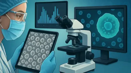
The limitations of manual counting paved the way for the stain-free revolution, which utilizes Digital Holographic Microscopy to analyze cells in their native state. Unlike traditional brightfield imaging that depends on light absorption and scattering to create contrast, this technology captures the phase shift of light as it passes through a specimen. This process creates quantitative phase images that reveal the intrinsic optical and morphological properties of cells without the need for any external chemical markers. Because the light interacts with the internal mass of the cell, the resulting data is a direct reflection of the cellular state rather than an artificial signal generated by a dye. This non-invasive approach allows researchers to observe cells exactly as they are in culture, preserving their physiological integrity for any future testing. By moving away from absorption-based imaging, laboratories have gained a far more detailed and objective view of their biological samples.
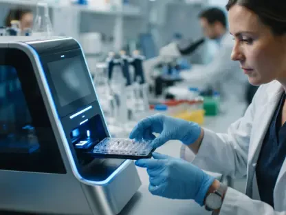
When this advanced imaging is paired with modern machine-learning algorithms, the system can distinguish between viable and non-viable cells by analyzing physical characteristics invisible to the human eye. This approach, utilized by advanced systems like the METTLER TOLEDO CytoDirect, eliminates the need for reagents entirely and automates the identification process. The software learns to recognize the specific holographic signatures of healthy cells versus those that are beginning to degrade, providing a level of accuracy and speed that was previously impossible. By removing the chemical variable from the equation, laboratories achieve a much higher level of data consistency. The integration of artificial intelligence ensures that every sample is analyzed using the same rigorous criteria, effectively removing the “noise” of human subjectivity. This synergy between physics and software has transformed cell counting from a manual craft into a high-precision digital science.
Beyond Binary Metrics: Navigating the Health Continuum
One of the most significant conceptual shifts enabled by stain-free technology is the move away from a binary live-or-dead classification of cell samples. Traditional stains only identify cells once they have reached a terminal state of membrane rupture, meaning that the data collected is often a lagging indicator of culture health. In contrast, analysis based on Digital Holographic Microscopy allows researchers to view cell viability as a health continuum. This means that subtle morphological changes, alterations in refractive index, or physiological stress can be detected long before a cell actually dies. By observing these early indicators, scientists can gain a much more nuanced understanding of how their cultures are responding to specific treatments or environmental conditions. This deeper insight allows for more precise experimental adjustments and a clearer picture of the underlying biological processes at play during a long-term study or production cycle.
By capturing these minute morphological details, stain-free counters provide a predictive capability that is invaluable for bioprocess optimization and quality control. Researchers can detect declining culture health early enough to intervene, potentially saving expensive batches of biological products that would have otherwise been lost. In fields like regenerative medicine and advanced biomanufacturing, this nuanced understanding of the cellular state is becoming a prerequisite for strict regulatory compliance. The ability to document the gradual progression of a cell population with quantitative data provides a robust audit trail for safety and efficacy. Rather than waiting for a culture to fail a simple dye-exclusion test, teams can now use holographic data to maintain optimal conditions throughout the entire growth cycle. This proactive approach to cell health management is essential for ensuring the high standards required for modern medical breakthroughs and biological products.
Operational Efficiency: Optimizing the Modern Workflow
Beyond the obvious scientific accuracy, the adoption of stain-free automated counting offers immediate operational benefits that improve overall laboratory productivity. By eliminating the need for reagent preparation, pipetting of hazardous dyes, and lengthy incubation times, researchers can move from a raw sample to a finalized result in a matter of seconds. This massive reduction in hands-on time allows staff to focus their expertise on high-level data interpretation and experimental design rather than the repetitive mechanics of counting. In a busy facility, the cumulative time saved across dozens of samples each day translates into faster project timelines and a significant reduction in labor costs. The simplicity of the “sample-in, results-out” workflow also lowers the barrier to entry for new staff, ensuring that even those with less experience can generate high-quality data that meets the rigorous standards of the organization.
The financial and logistical advantages of this technology are equally compelling for long-term facility management. Removing the recurring costs associated with staining reagents and specialized consumables provides a substantial economic benefit for high-volume laboratories. Furthermore, because the cells remain unstained and unharmed during the counting process, they can be returned to the culture or used for subsequent procedures like genomic sequencing or flow cytometry. This preservation of material is vital for sensitive workflows where every single cell is a precious resource, such as in personalized medicine or rare disease research. By maximizing the utility of every sample, labs can reduce the total volume of starting material required for an experiment, further lowering costs and ethical concerns. This streamlined approach to resource management ensures that the laboratory remains both lean and highly effective in its pursuit of scientific discovery.
Strategic Implementation: Real-World Evidence and Next Steps
The shift to stain-free technology has already produced tangible results in both academic and industrial settings across the globe. Institutions that integrated these systems reported a significant reduction in total analysis time and a marked improvement in the overall efficiency of their experimental pipelines. Researchers at the University of Bern, for example, successfully simplified their daily cell culture tasks by removing the staining step entirely, allowing them to process more samples with greater confidence in their data. Early-career scientists found that the removal of manual intervention allowed them to dedicate more time to complex problem-solving and less to the rote tasks that previously dominated their mornings. These successes demonstrated that the transition to automated, label-free imaging was not merely a technological upgrade but a fundamental improvement in how biological research is conducted on a daily basis.
To capitalize on these advancements, laboratories were encouraged to audit their existing counting protocols and identify the hidden costs of manual variability and reagent waste. The transition required a strategic shift in how teams valued their time and their samples, moving toward a model where data integrity was prioritized from the very first step of the workflow. Organizations that successfully adopted these platforms found that they were better positioned to meet the demands of advanced biomanufacturing and personalized medicine. Moving forward, the integration of these high-fidelity data streams into digital management systems will become the standard for any facility aiming to remain competitive. By embracing the synergy of holography and artificial intelligence, the scientific community has ensured that the foundational step of cell counting is now as sophisticated and reliable as the advanced therapies it was designed to support.