
The landscape of oncology is shifting from a generalized “one-size-fits-all” approach to a molecularly granular understanding of how individual tumors behave. Ivan Kairatov, a prominent Biopharma expert with a distinguished background in research and development, has spent his career at the intersection of technological innovation and clinical application. His work focuses on how emerging tools can transform massive datasets into actionable insights for patients facing the most challenging diagnoses. In this discussion, we explore the revolutionary potential of single-cell analysis, specifically a new machine learning framework designed to decode the complex cellular architecture of cancer to predict patient survival with unprecedented accuracy.
The conversation touches on the limitations of traditional bulk sequencing, which often obscures critical biological signals by averaging data across a tumor. We delve into the mechanics of the scSurvival model, examining how it uses weight-based filtering to isolate high-risk cell populations from more than 150 clinical cases. Furthermore, we examine the practical implications of these findings for diseases like melanoma and liver cancer, where identifying specific immune patterns can dictate the success of immunotherapy. Ultimately, the discussion highlights the shift from simply identifying risk to understanding the underlying cellular “why” that governs disease progression.
Conventional methods often average cell data across a tumor, essentially putting the biological picture in a blender. How does this loss of nuance impact our understanding of disease progression, and what specific risks are overlooked when we fail to see the finer details of individual cells?
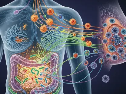
When we rely on traditional methods, we are essentially looking at a blurred snapshot of a highly complex and dynamic environment. Every tumor is a unique mosaic of cells, and by “blending” them together, we lose the ability to see the specific outliers that might be driving a patient’s relapse or resistance to treatment. This loss of nuance means we often miss rare but aggressive cell populations that constitute only a small fraction of the tumor but dictate the ultimate survival outcome. For years, researchers have collected single-cell gene expression data from thousands to millions of tumor cells, yet the tools to interpret that data without losing the finer details were lacking. By ignoring these individual variations, we risk undertreating patients who harbor high-risk cellular signatures or, conversely, over-treating those whose primary tumor mass appears aggressive but lacks the specific cell-level drivers of progression. It is a frustrating reality where the “big picture” actually hides the most critical biological truths needed to save lives.
The scSurvival model assigns specific weights to individual cells based on their relationship to patient survival. Could you walk through the technical steps of how this filtering process works and how it manages to distinguish high-risk cell populations from less significant data?
The technical brilliance of scSurvival lies in its ability to take a “fine-tooth comb” to massive datasets, which was a primary goal for the team at Oregon Health & Science University. To accomplish this, the model assigns each individual cell a specific weight based on the mathematical degree to which that cell’s expression profile is statistically related to patient survival. This filtering process is vital because it allows the algorithm to essentially ignore the “noise” generated by less important cells that don’t influence the disease’s trajectory. Once these weights are established, the model averages the data only from the weighted cells, creating a refined basis for its survival predictions rather than a generic average. By training on single-cell datasets paired with survival data from hundreds of clinical cases, the framework learns to recognize which specific biological patterns are the true harbingers of risk. It is a highly targeted approach that ensures the most influential biological actors within the tumor are the ones guiding the clinical prognosis.
Testing on melanoma and liver cancer cohorts has shown more accurate outcomes than traditional methods. What specific immune or tumor cell patterns were identified in these high-risk patients, and how might these findings change the way doctors approach immunotherapy responses?
The clinical application of this model on more than 150 cancer patients has yielded some of the most exciting data we have seen in single-cell oncology. In the melanoma cohorts, the model successfully traced survival predictions back to specific immune and tumor cell groups that were directly linked to how a patient responded to immunotherapy. This is a game-changer because it allows us to identify the presence or absence of specific cell populations that act as “gatekeepers” for treatment success before a patient even starts a regimen. In liver cancer, the tool identified cell-level patterns that shaped tumor behavior in ways that traditional bulk sequencing simply could not detect. For a doctor, having this level of detail means they can move beyond trial and error, choosing therapies that are specifically calibrated to the cellular landscape of that individual’s cancer. It transforms immunotherapy from a broad-spectrum hope into a precision-guided strike based on the actual cellular makeup of the patient’s unique mosaic.
Every tumor presents a unique mosaic of cells that dictate how the disease will progress. In a clinical setting, how would a tool that identifies the “why” behind a patient’s risk profile assist oncologists in personalizing treatment plans for difficult-to-treat cancers?
In the high-stakes environment of oncology, knowing that a patient is at high risk is only half the battle; the real power lies in understanding the biological “clues” as to why that risk exists. As noted by experts at the National Cancer Institute, a tool that provides the “why” behind a risk profile is indispensable for managing difficult-to-treat cancers where standard protocols often fail. If an oncologist can see that a patient’s high-risk status is driven by a specific cluster of immune-evading cells, they can pivot to a treatment plan that specifically targets that evasion tactic. This level of insight allows for the creation of a truly personalized roadmap, where every therapeutic decision is backed by the granular data of the tumor’s internal architecture. It replaces the anxiety of the unknown with a clear, data-driven strategy, potentially sparing patients from the side effects of ineffective treatments while accelerating the use of those that will actually work. This research, supported by multiple NIH grants including R01CA283171 and U01CA253472, proves that we are finally moving toward a future where the “mosaic” of the tumor is a map rather than a mystery.
What is your forecast for AI-driven single-cell analysis in oncology?
My forecast is that within the next decade, single-cell analysis driven by machine learning will become the standard of care, moving from specialized research labs into every major diagnostic clinic. We will see models like scSurvival integrated into real-time diagnostic workflows, where a biopsy can be sequenced and analyzed within days to provide a survival and response prediction before the first treatment is even administered. The era of “blender biology” is coming to a definitive end, replaced by a “high-resolution” era where we treat the specific cells causing the harm rather than the organ as a whole. I expect we will see a surge in the discovery of rare cell types that we never knew were critical to cancer progression, leading to a new wave of drug development targeting these newly identified high-risk populations. Ultimately, this technology will bridge the gap between massive genomic datasets and the individual patient experience, making “precision medicine” a reality for millions of people rather than just a buzzword.