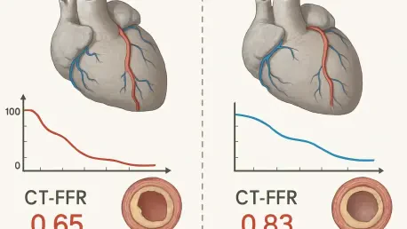

For decades, cardiologists have sought a definitive non-invasive method to measure blood flow through coronary arteries without resorting to risky catheterization procedures, and computed tomography-derived fractional flow reserve (CT-FFR) emerged as a transformative technology. However, its accuracy remains contingent on several physiological variables that fluctuate during a single heartbeat. A recent study published in BMC Medical Imaging has brought this technical dilemma to the forefront, examining how the specific phases of the cardiac cycle—systole and diastole—influence the diagnostic precision of these virtual assessments. By analyzing the impact of cardiac phase selection, researchers have provided critical insights into whether the industry-standard reliance on the diastolic phase is truly the most effective approach for all patients. This investigation is particularly relevant today in 2026, as clinicians strive to refine imaging protocols to account for anatomical complexities like myocardial bridging and high heart rates during cardiac scans.
Defining Study Scope and Research Methodology
The core of this retrospective diagnostic-accuracy study centered on a detailed analysis of 138 patients, comprising 155 individual coronary vessels, all imaged using a state-of-the-art 320-detector-row CT platform. Unlike previous studies that might have accepted standardized defaults, this research team utilized a steady-state computational framework to calculate CT-FFR from both systolic and diastolic reconstructions independently. This dual-phase approach allowed for a direct head-to-head comparison against the “gold standard” of invasive fractional flow reserve measurements obtained in a catheterization lab. By setting a strict hemodynamic threshold of 0.80 or less to define significant ischemia, the researchers could evaluate how well each cardiac phase predicted the actual physiological state of the vessel. The methodology was further strengthened by the application of patient-clustered bootstrap statistical modeling, ensuring that the findings remained robust even when multiple vessels from the same patient were analyzed.
Traditionally, the medical community has favored diastolic-phase data because it generally offers the best image quality with the least amount of motion blur, which is essential for accurate lumen segmentation. However, the presence of myocardial bridging—a condition where a segment of a coronary artery is tunneled through the heart muscle—creates a unique physiological challenge that might favor systolic imaging. During systole, the contraction of the myocardium can compress these bridged segments, potentially altering the flow dynamics in a way that diastolic imaging might overlook. Furthermore, the variability of patient heart rates introduces another layer of complexity; higher rates often shorten the diastolic window, leading some to speculate that the systolic phase might provide a more stable alternative. This research sought to demystify these assumptions by providing empirical evidence on how these specific anatomical and physiological factors influence final CT-FFR values across the cardiac cycle.
Analyzing Performance and Clinical Implications
Upon analyzing the data, a clear trend emerged regarding the overall discriminatory power of the two phases, with the diastolic phase maintaining a distinct statistical lead. The Area Under the Receiver Operating Characteristic curve, a standard measure of diagnostic performance, was measured at 0.91 for diastolic reconstructions compared to 0.87 for systolic datasets. This difference of 0.039 proved to be statistically significant, reinforcing the notion that the diastolic phase remains the more reliable foundation for general physiological assessment in a clinical setting. This finding suggests that for the average patient, the clearer visualization of the coronary anatomy during the heart’s relaxed state provides a more accurate map for computational fluid dynamics. While systolic imaging remains a technically viable option, it consistently trailed in its ability to differentiate between hemodynamically significant stenosis and non-obstructive lesions across the broader patient population during the clinical assessment.
The most compelling findings were revealed during post hoc analyses of complex diagnostic scenarios, specifically focusing on the invasive FFR “gray zone” between 0.75 and 0.85. In these borderline cases, where clinical decisions are most difficult to make, the diastolic phase outperformed the systolic phase by a substantial margin of 21.6 percentage points. Similarly, in the presence of myocardial bridging, the diastolic phase demonstrated its resilience as a diagnostic tool, maintaining a high AUC of 0.90 while the systolic phase dropped to 0.79. This suggests that the presence of bridging actually amplifies the performance gap between the phases, confirming that the diastolic approach should remain the primary choice for patients with complex coronary anatomy. Furthermore, the study did not find a significant difference in phase performance when patients were stratified by heart rate, suggesting that the diastolic phase remains the reliable standard even in patients with higher pulse rates.
Looking ahead to the further refinement of cardiovascular imaging, this study established that while the systolic phase was technically feasible, it provided no distinct advantage over traditional diastolic assessments. Medical facilities should, therefore, continue to prioritize high-quality diastolic image acquisition as the foundation for CT-FFR calculations, particularly for patients presenting with borderline stenosis or suspected myocardial bridging. Future developments in imaging software might integrate these findings by automatically flagging cases in the diagnostic “gray zone” for more rigorous diastolic review. Moreover, the discovery that heart rate did not significantly shift the balance in favor of systolic imaging simplified protocol standards for 2026 and beyond. By focusing on optimizing diastolic windows, even in patients with elevated pulses, healthcare providers can ensure higher diagnostic accuracy and reduce the necessity for unnecessary invasive interventions. The research ultimately validated the current clinical preference for diastolic datasets.