
Ivan Kairatov is a distinguished biopharma expert whose career has been defined by a relentless pursuit of innovation within the research and development sector. With deep-seated expertise in bridging the gap between complex biotechnological advancements and clinical applications, he has become a leading voice in how data-driven insights can revolutionize patient care. In this discussion, we explore a significant breakthrough in neurosurgery—the use of geometric markers to treat a condition that often masquerades as permanent cognitive decline.
Our conversation covers the persistent challenges of diagnosing idiopathic normal pressure hydrocephalus (iNPH) and the limitations of traditional imaging techniques that frequently lead to unsuccessful surgical interventions. We examine the sophisticated shift from simple linear measurements to 3D shape analysis, specifically the role of “asphericity” in predicting surgical success. Furthermore, we discuss the practical hurdles hospitals face when integrating machine learning into standard neuroimaging protocols and how these geometric insights could eventually serve as a blueprint for treating a wide array of complex neurological conditions.
Idiopathic normal pressure hydrocephalus symptoms like gait instability and cognitive decline often mimic other dementias, leading to frequent underdiagnosis. What specific hurdles do clinicians encounter during the diagnostic process, and how does the current lack of predictive markers impact the rate of ineffective shunt surgeries?
The diagnostic landscape for iNPH is incredibly treacherous because its hallmark symptoms—abnormal gait, urinary urgency or incontinence, and cognitive decline—overlap almost perfectly with more common conditions like Alzheimer’s or Parkinson’s disease. Clinicians often find themselves in a high-stakes guessing game where the physical evidence of enlarged brain ventricles doesn’t always correlate with the clinical reality of the patient’s neurological health. Because our current diagnostic tools lack precision, we are often unable to distinguish between a patient who has a treatable fluid buildup and one whose brain structure has simply changed due to irreversible neurodegeneration. This lack of predictive clarity is devastating in a surgical context; without reliable markers, a significant number of elderly patients undergo invasive shunt procedures that ultimately offer no relief. The weight of an ineffective surgery is not just a clinical failure—it represents a period of physical trauma and dashed hopes for families who believed they were on the cusp of reversing their loved one’s dementia.
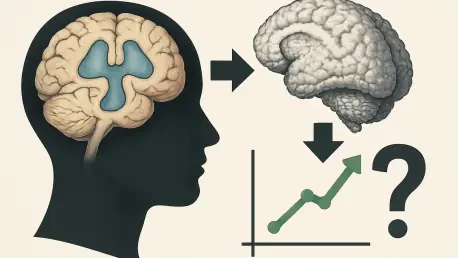
Quantifying 3D geometric features, specifically asphericity, has shown promise in predicting patient outcomes for those with enlarged brain ventricles. How does this shape-based analysis differ from traditional linear measurements, and what is the step-by-step process for applying machine learning to these neuroimaging scans?
Traditional neuroimaging has historically relied on linear measurements—essentially taking a ruler to a two-dimensional slice of a brain scan—which provides a very limited, static view of a highly complex, volumetric structure. Shape-based analysis, particularly the study of asphericity, moves beyond mere size to examine how much the lateral ventricles deviate from a perfect sphere, capturing the subtle distortions caused by fluid pressure. In the study involving 170 patients with iNPH, researchers utilized advanced 3D imaging to map the entire volume of these ventricles, allowing for a much more nuanced understanding of the brain’s internal architecture. The machine learning process begins by feeding these high-resolution 3D scans into an algorithm that has been trained to recognize specific geometric patterns associated with positive surgical outcomes. By quantifying these features across a large cohort, the system can identify “asphericity” as a key indicator, providing a statistical probability of success that a human eye simply cannot discern from a standard MRI.
Shunt surgery offers a rare opportunity to reverse dementia-like symptoms, yet selecting the right candidates remains difficult. Beyond asphericity, what clinical metrics do you prioritize when evaluating surgical viability, and how could these geometric insights help spare patients from the risks of invasive, non-beneficial procedures?
When we evaluate a patient for a shunt, we are looking for a very specific “win” where the drainage of excess cerebrospinal fluid will actually restore function, but we must balance this against the inherent risks of brain surgery in an older population. Beyond the promising data on asphericity, we look closely at the severity of gait disturbance and the timing of symptom onset, as early intervention is often the difference between full recovery and permanent disability. The introduction of 3D geometric insights acts as a critical filter, allowing us to identify those “non-responders” before they ever reach the operating table. By using these markers to deselect patients who are statistically unlikely to improve, we protect them from the potential complications of surgery, such as infections or hemorrhages, which can be catastrophic for someone already in a fragile cognitive state. It is about moving toward a model of “precision neurosurgery,” where every incision is backed by a high degree of mathematical certainty that the patient’s quality of life will actually improve.
Implementing advanced 3D imaging tools into standard clinical practice involves technical and logistical shifts. What infrastructure is necessary for hospitals to adopt this ventricle analysis, and how do you envision this technology evolving to help patients with other types of complex brain conditions?
To move this technology from a research environment into a busy hospital setting, we need a robust digital infrastructure that includes high-performance computing capabilities and seamless integration with existing Picture Archiving and Communication Systems (PACS). Radiologists and neurologists will need specialized software interfaces that can automate the 3D segmentation of the ventricles, turning raw MRI data into a clear “asphericity score” without adding hours to the diagnostic workflow. Looking forward, the evolution of this technology is incredibly exciting; the same principles of geometric quantification could be applied to track the progression of multiple sclerosis or to map the subtle structural shifts in early-onset Alzheimer’s. If we can master the art of reading the brain’s “topography” through machine learning, we can begin to detect pathological changes years before the first physical symptoms even emerge.
What is your forecast for the use of geometric brain markers in neurosurgery?
I believe that within the next decade, geometric brain markers will become the gold standard for surgical planning, effectively ending the era of “wait and see” diagnostics for fluid-related brain disorders. As our datasets grow beyond the initial 170 patients and encompass more diverse populations, these 3D markers will be integrated into real-time surgical navigation systems, allowing surgeons to visualize not just where to place a shunt, but exactly how much pressure change is required for that specific brain’s geometry. We are moving toward a future where “dementia” is no longer seen as a monolithic, irreversible sentence, but as a collection of specific physical conditions—some of which can be mapped, measured, and corrected with mathematical precision. This shift will fundamentally change the economics of neurosurgery by reducing the rate of failed procedures and, more importantly, it will offer a definitive roadmap for recovery to thousands of patients who are currently lost in the diagnostic gap.