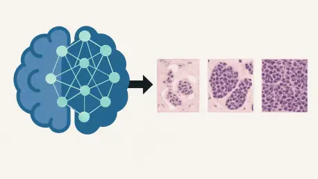

Transformer models utilize self-attention mechanisms to weigh the importance of different tumor regions, capturing global structural context better than traditional convolutional networks. In the current medical landscape of 2026, the convergence of oncology and artificial intelligence has yielded diagnostic tools that significantly reduce the reliance on invasive procedures for tumor characterization. The Ki-67 protein serves as a primary biological marker for cellular proliferation, acting as a microscopic clock that measures how aggressively a cancer cell is dividing and spreading. Traditionally, determining the Ki-67 index requires a physical biopsy, where a needle extracts tissue for immunohistochemical staining. However, breast cancer is notoriously heterogeneous, meaning that a single tissue sample may not accurately represent the biological state of the entire tumor mass. This sampling error can lead to a misleading prognosis, potentially resulting in inappropriate treatment plans for patients. By utilizing Dynamic Contrast-Enhanced Magnetic Resonance Imaging (DCE-MRI), modern researchers are developing a “virtual biopsy” approach. This methodology evaluates the entire tumor volume preoperatively, offering a more comprehensive diagnostic picture that accounts for the varying degrees of malignancy across different regions of the mass. Through this lens, deep learning is not just assisting radiologists but is fundamentally redefining how we quantify tumor biology in a non-invasive manner.
Data Standardization: Ensuring Consistency in Imaging Analysis
To ensure that artificial intelligence can provide reliable results across different medical institutions, a rigorous data preprocessing pipeline is essential. Recent large-scale investigations involving over 300 patients across multiple hospital sites have highlighted the necessity of standardizing images before they enter a neural network. Because MRI scans are produced by machines from various manufacturers, such as GE and SIEMENS, the raw images often contain subtle differences in resolution and signal intensity that could confuse an AI model. To mitigate these variations, researchers now resample all images to a uniform one-millimeter isotropic voxel size. This ensures that every pixel represents the same physical space regardless of the original scanner’s settings. Furthermore, techniques like N4 bias field correction are employed to remove magnetic field inhomogeneities, which can cause artificial brightness variations. By cleaning the data in this manner, the deep learning model can focus exclusively on the biological features of the tumor rather than the technical artifacts of the imaging equipment.
Moving beyond simple image cleanup, the adoption of a 2.5-dimensional processing strategy has proven to be a highly efficient way to represent three-dimensional tumor structures. Instead of analyzing a single flat image or attempting to process an entire 3D volume, which requires immense computational power, researchers extract eleven critical slices centered on the largest cross-section of the tumor. This approach captures the essential depth and structural variety of the lesion without the overhead of full 3D modeling. Once these slices are selected, they are fed into a specialized Convolutional Neural Network known as ResNet-50. This architecture is renowned for its use of residual blocks, which allow the network to learn complex patterns without the degradation of information that often occurs in deeper models. By removing the final classification layer of ResNet-50, the system is transformed into a high-powered feature extractor. This process converts every visual slice of the tumor into a 2048-dimensional mathematical fingerprint, creating a dense numerical representation of the tumor’s texture, shape, and vascularity.
Architectural Evolution: Multi-Instance Learning Versus Transformers
Once the visual data is converted into numerical vectors, the primary challenge becomes how to aggregate this information to make a final prediction for the patient. One effective strategy is Multi-Instance Learning, which treats the collection of tumor slices as a “bag” of individual instances. This framework looks for the distribution and frequency of specific visual patterns across the entire tumor. For example, by using a Predictive Likelihood Histogram, the model can determine if certain aggressive characteristics appear frequently enough to warrant a high Ki-67 classification. Another technique within this framework, the Bag-of-Words model, treats specific visual features as “words” and counts their occurrence to build a profile of the tumor. Recent tests have shown that these Multi-Instance Learning methods are highly robust, achieving an impressive Area Under the Curve of 0.825 during validation trials. This capability allows doctors to see a holistic view of the tumor’s potential for growth based on the most suspicious regions identified by the algorithm.
While Multi-Instance Learning focuses on the frequency of features, the shift toward Transformer-based fusion models has introduced a new level of sophistication by focusing on the relationships between different parts of the tumor. Using a self-attention mechanism, the Transformer calculates how much weight or importance one specific slice of an MRI should have relative to all others. This allows the model to understand the global context of the tumor’s structure, recognizing that a feature on the outer margin may be more significant when viewed in relation to a necrotic area in the center. In head-to-head comparisons, the Transformer model has emerged as the superior diagnostic tool, reaching a peak accuracy of nearly 84 percent. This suggests that the spatial relationship and the “conversation” between different tumor regions contain vital biological clues that simpler models might overlook. The ability of Transformers to weigh these complex interactions effectively mimics the synthesis of information performed by the most experienced human pathologists.
Performance Metrics: Surpassing Radiomics and Clinical Benchmarks
To validate the efficacy of these advanced neural networks, they must be benchmarked against traditional diagnostic methods, such as radiomics and clinical data analysis. Radiomics involves the manual extraction of over a thousand quantitative features, including tumor shape, intensity, and texture, which are then analyzed using statistical regression. While this was once the gold standard for computational oncology, recent findings indicate that these “hand-crafted” features are often too simple to capture the nuanced biological signals of Ki-67 expression. In comparative studies, radiomic models achieved an accuracy significantly lower than their deep learning counterparts, illustrating a clear gap between human-defined metrics and the autonomous feature learning capabilities of modern AI. The complexity of cancer biology appears to require the multi-layered analysis that only deep learning can provide, as the most critical indicators of proliferation are often invisible to traditional mathematical formulas.
Further analysis has revealed that standard clinical factors, such as a patient’s age, body mass index, or menopausal status, provide very little predictive value for Ki-67 levels. Interestingly, among all standard clinical markers, only HER-2 status showed a notable correlation with tumor proliferation. This highlights a significant limitation in current clinical practice: many of the data points traditionally used to assess risk are disconnected from the actual molecular behavior of the cancer. The failure of demographic data to predict aggressiveness reinforces the importance of advanced imaging analysis. By looking directly at the tissue’s vascular patterns and structural integrity through DCE-MRI, deep learning models can find the specific signatures of rapid cell division that are entirely absent from a patient’s medical history or basic radiological report. This evidence strongly supports the integration of AI-driven imaging tools as a necessary supplement to, and perhaps eventually a replacement for, traditional clinical risk assessments.
Clinical Interpretability: Seeing through the Artificial Intelligence Lens
A persistent challenge in the adoption of medical AI is the “black box” problem, where the reasoning behind a machine’s decision remains hidden from the clinician. To address this, researchers have implemented Gradient-weighted Class Activation Mapping, which generates heatmaps that overlay directly onto the original MRI scans. These visualizations show exactly which areas of the tumor the model prioritized when determining the Ki-67 level. It was discovered that the Transformer model does not simply look at the center of the tumor mass; instead, it focuses on the active parenchymal regions and the interface where the tumor meets healthy breast tissue. These findings are highly significant because they align with biological reality—the margins of a tumor are often the most active zones of cellular division and invasion. Providing this visual evidence allows radiologists to trust the AI’s output, as they can verify that the model is making decisions based on relevant anatomical features rather than noise or artifacts.
The transition from manual feature engineering to autonomous feature learning represents a paradigm shift in how we understand medical imaging. By using visualization tools, researchers have confirmed that these models are identifying complex biological phenomena, such as internal necrosis or irregular vascular growth, which are known indicators of high-grade malignancy. This level of transparency is crucial for the eventual approval of these tools by regulatory bodies and for their daily use in hospitals. The stability of these models across different medical centers also suggests that the features being learned are universal biological markers rather than site-specific patterns. As these tools become more refined, they provide a bridge between the digital world of data science and the physical reality of patient care, ensuring that the insights gained from deep learning are both scientifically sound and clinically actionable for the medical community.
Clinical Trajectory: Shaping the Future of Precision Oncology
Despite the current successes in predicting Ki-67 levels, several avenues for future enhancement remain open as the industry moves toward 2027 and beyond. Current models often rely on a single-phase MRI, but cancer is a dynamic process that changes over time. Future iterations of this technology could incorporate multiple temporal phases of contrast enhancement, allowing the AI to track how blood flows into and out of a tumor over several minutes. This hemodynamic data could provide an even deeper look into the metabolic activity of the cancer cells. Additionally, the development of hybrid architectures that combine the local sensitivity of Multi-Instance Learning with the global contextual awareness of Transformers could push predictive precision to even higher levels. As computational power continues to increase, the possibility of moving from 2.5-dimensional slices to fully integrated 3D volumetric analysis will likely become the new standard for oncological imaging.
The study successfully demonstrated that deep learning could effectively bridge the gap between non-invasive imaging and molecular pathology. By accurately identifying Ki-67 levels, the technology held the potential to refine treatment paths, allowing clinicians to distinguish between patients who required immediate, aggressive chemotherapy and those who could safely pursue less toxic alternatives. The researchers concluded that the use of a Transformer-based fusion model provided the most stable and accurate results, outperforming both traditional radiomics and standard clinical assessments. These advancements suggested a future where the “virtual biopsy” became a routine part of preoperative planning, reducing the physical burden on patients while increasing the precision of their care. Ultimately, the integration of these digital tools into the clinical workflow represented a major step forward in the quest for personalized medicine, ensuring that every therapeutic decision was grounded in a complete understanding of the tumor’s unique biological profile.