
The blue residues found on the walls of the tombs in the Valley of the Kings have unexpectedly become the foundation for a sophisticated leap in medical diagnostics. Known as Egyptian blue, this pigment was initially synthesized roughly 4,500 years ago, marking a pivotal moment in the history of human chemistry as one of the first artificial pigments ever created. While it once served to immortalize pharaohs and signify the divine, contemporary research has reimagined it as a cutting-edge nanomaterial. Scientists are now exfoliating this ancient substance into layers so thin that they exist at the atomic scale, enabling a level of precision in imaging that was previously unthinkable. This bridge between antiquity and the present showcases how historical craftsmanship can inform modern healthcare solutions. By repurposing this enduring material, researchers are solving complex biological visualization problems, proving that ancient innovations still hold secrets for the digital age.
Historical Origins: From Ancient Kilns to Modern Laboratories
Calcium copper silicate, or cuprorivaite, represents the pinnacle of ancient material science, created by artisans who lacked modern labs but possessed deep practical knowledge of mineralogy. They discovered that heating a specific mixture of limestone, sand, and copper-rich minerals—such as malachite or azurite—at temperatures exceeding 850 degrees Celsius produced a vibrant blue glass-like substance. This process required precise control over the kiln environment to avoid turning the mixture into a black sludge or an inconsistent green. The resulting pigment was not merely a dye but a crystalline structure that offered unprecedented durability against sunlight and moisture. Because lapis lazuli was a prohibitively expensive import from distant lands, this synthetic alternative democratized the use of the color blue in royal and religious art. Its widespread adoption across the Mediterranean ensured that the material would survive for millennia, eventually becoming a point of interest for researchers.
The chemical robustness of Egyptian blue is a testament to the sophistication of early chemical engineering, as the pigment remains vibrant even after thousands of years of exposure to the elements. Unlike organic dyes derived from plants or insects, which degrade under ultraviolet light or through oxidation, this mineral-based compound maintains its structural integrity. This longevity is what initially caught the eye of material scientists, who began to investigate the unique molecular lattice of the substance. They realized that the stability of the copper ions within the silicate framework was not just aesthetically pleasing but also offered specific physical properties that could be exploited in electronic and optical applications. This shift from art history to material science underscores a growing trend where researchers look backward to find stable, non-toxic materials for high-tech use. The enduring nature of the pigment ensures that any diagnostic tool derived from it will possess a reliability that exceeds organic alternatives.
Technical Superiority: Nanoscale Imaging and Signal Stability
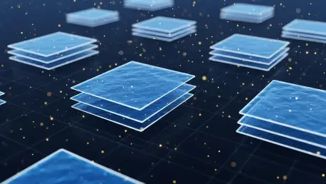
The transition of Egyptian blue from a bulk pigment to a high-tech imaging agent relies on a process known as liquid-phase exfoliation, which breaks the mineral down into nanosheets. These individual layers are exceptionally thin, often measuring only a few nanometers in height, which allows them to integrate into biological systems without causing mechanical disruption. At this scale, the pigment exhibits a property called near-infrared fluorescence, where it absorbs energy and re-emits it at wavelengths beyond the visible spectrum. This particular range of light is highly advantageous for medical imaging because it falls within the optical window of human tissue. Standard visible light is frequently scattered by cellular structures or absorbed by hemoglobin, creating a blurred image that lacks depth. In contrast, the near-infrared emissions from Egyptian blue can pass through several centimeters of flesh with minimal interference, providing a clear path for non-invasive mapping.
One of the most persistent challenges in fluorescent imaging is the phenomenon of photobleaching, where a marker loses its ability to glow after repeated exposure to light. Most modern organic dyes suffer from this degradation, which limits the duration of time a scientist can track a biological event. Egyptian blue, however, displays a remarkable resistance to this process due to its inorganic, crystalline nature. This means that a single dose can provide a constant signal over hours or days, which is essential for studying long-term cellular developments or drug interactions. This stability allows for continuous monitoring of a subject without the need for repeated injections. In a laboratory setting, this translates to more reliable data and a reduction in the variables that can skew results. The pigment’s ability to stay on under intense illumination sets it apart from almost every other synthetic fluorophore, offering an exceptional signal-to-noise ratio that stands out clearly.
Empirical Validation: Clinical Outcomes and Future Protocols
To move the technology from a theoretical concept to a practical medical tool, extensive testing was conducted on various biological models. In these experiments, the nanosheets were integrated into the embryos of fruit flies, allowing researchers to track the intricate dance of cell division during early development. The pigment demonstrated excellent biocompatibility, as the embryos continued to grow into healthy adults without side effects. Similarly, when applied to plant systems, the near-infrared emissions were able to penetrate chlorophyll-rich layers with ease, mapping nutrient distribution in a way that was previously impossible. These tests confirmed that the nanosheets did not aggregate within cells, ensuring they remained evenly distributed to provide a consistent visual map. The success of these trials across different kingdoms of life suggested that the pigment was a universal imaging agent, capable of functioning in environments with high levels of interference and barriers.
The journey of Egyptian blue from a decorative glaze to a sophisticated diagnostic tool demonstrated the value of re-examining ancient materials through the lens of modern physics. It became clear that the stability of this mineral offered solutions to the most persistent problems in oncology, where the pigment served as a guide for tumor removal. By coating nanosheets with ligands that targeted cancer markers, medical teams “painted” tumors with a glow that allowed surgeons to see exact boundaries. This precision reduced recurrence rates and improved patient recovery. Moving forward, researchers prioritized the scaling of production to meet the global demand for effective imaging agents. They established safety protocols that allowed for the transition from experimental use to clinical standards. By bridging historical ingenuity and contemporary needs, the medical community secured a powerful method for visualizing life, creating a blueprint for discovering other ancient substances for modern use.