
The traditional methodology of identifying genetic mutations in cancer patients often relies on time-consuming processes that destroy precious biopsy samples before a treatment plan can even begin. This fundamental conflict between diagnostic necessity and sample preservation has led to the emergence of AI-enhanced fluorescence lifetime imaging (FLIM) as a pivotal solution in 2026. By shifting the focus from chemical-heavy sequencing to non-invasive optical sensing, this technology addresses the critical “tissue scarcity” problem. Modern oncological screening now identifies tumors at much earlier stages, resulting in smaller biopsies that cannot withstand the destructive nature of conventional genomic testing.
Introduction to AI-Enhanced Fluorescence Lifetime Imaging
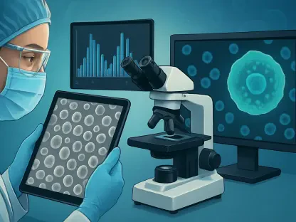
Fluorescence lifetime imaging operates on the principle that natural molecules within biological tissue emit light for a specific duration after being excited by a laser. Unlike traditional imaging that only measures the intensity of light, FLIM records the decay rate of this fluorescence, which serves as a highly sensitive indicator of the cellular microenvironment. This approach is inherently label-free, meaning it does not require the application of external dyes or contrast agents that can alter or damage the sample.
Integrating machine learning into this optical process has transformed raw decay data into a predictive diagnostic tool. By training algorithms on vast datasets of known genetic profiles, researchers have enabled the system to “read” the metabolic signatures associated with specific pathologies. This synergy between optics and computation provides a window into the molecular state of a patient’s cells without the need for invasive chemical disruption, positioning it as a cornerstone of modern personalized medicine.
Technical Architecture and Core Capabilities
Label-Free Optical Sensing and Signal Acquisition
The primary strength of FLIM lies in its ability to capture the metabolic state of cells by analyzing the nanosecond-scale duration of light emission. While intensity-based imaging can be affected by the concentration of fluorophores or the thickness of the tissue, the fluorescence lifetime is an intrinsic property that remains independent of these variables. This allows for a much more objective assessment of biological samples, revealing shifts in metabolic co-factors like NADH and FAD that are often the first indicators of oncogenic transformation.
This sensing process is entirely non-destructive, which is a significant departure from the standard protocols of molecular pathology. Because the integrity of the biological sample is maintained, clinicians can use the same biopsy for multiple types of analysis if necessary. This capability is vital in scenarios where obtaining a second biopsy is impossible or carries significant risk to the patient, ensuring that every millimeter of extracted tissue provides the maximum possible diagnostic value.
Machine Learning Frameworks for Mutation Prediction
At the heart of the predictive capability is a sophisticated deep learning framework designed to recognize patterns that remain invisible to the human eye. These models analyze the complex, multi-exponential decay curves generated by FLIM to identify specific genetic markers, such as mutations in the epidermal growth factor receptor (EGFR). By correlating the precise timing of fluorescence with genomic data, the AI can distinguish between healthy tissue and various mutation subtypes with remarkable sensitivity.
Training these models requires a rigorous process of feature extraction, where the algorithm learns to filter out background noise while emphasizing the subtle optical signatures of diseased cells. This allows the system to achieve a high degree of specificity, ensuring that clinicians can rely on the results to make treatment decisions. Moreover, as the database of optical profiles grows, the accuracy of these predictions continues to refine, making the system more robust against the natural variability found in human tissue.
Recent Advancements in Optical Genetic Profiling
The field has recently experienced a major shift toward real-time diagnostics, moving away from the weeks-long wait times associated with traditional sequencing. Hardware miniaturization has enabled the development of compact FLIM devices that can be deployed directly within the clinical environment. This transition to “point-of-care” testing means that genetic information can be obtained almost immediately after a biopsy is taken, bypassing the logistical delays of centralized laboratory processing.
Furthermore, the convergence of high-speed optics and automated AI analysis is influencing the broader trajectory of molecular biology. We are seeing a move toward holistic diagnostic platforms that do not just look for a single mutation but provide a comprehensive overview of the tissue’s molecular landscape. This advancement suggests a future where the distinction between imaging and genetic sequencing becomes increasingly blurred, leading to more integrated and efficient workflows in the pathology department.
Clinical Applications in Precision Oncology
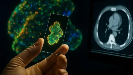
Real-world deployment of this technology has already demonstrated its transformative potential, particularly in the management of lung cancer at major medical institutions. In environments like the University of Edinburgh and NHS Lothian, FLIM-AI platforms have been used to identify patient eligibility for targeted therapies in a fraction of the time required by previous methods. This speed is life-saving, as it allows patients to start effective treatments within minutes of their diagnostic procedure rather than waiting for fourteen to twenty-one days for a lab report.
The technology is especially effective in preserving limited biopsy material obtained from early-stage screenings. In these cases, the sample size is often too small for multiple tests; however, because the AI-enhanced imaging is non-destructive, it leaves the tissue available for further validation or auxiliary testing. This application ensures that even the most vulnerable patients, who might only have a single opportunity for a successful biopsy, receive the benefit of comprehensive molecular profiling.
Barriers to Widespread Clinical Implementation
Despite its promise, the technology faces significant hurdles regarding the standardization of AI models across various imaging hardware. Because different microscopes and sensors can produce slightly different optical outputs, creating a “universal” algorithm remains a complex technical challenge. Without strict calibration protocols, the performance of the system could vary between hospitals, which complicates the process of achieving widespread regulatory approval from agencies like the FDA or EMA.
Moreover, there are physical limitations to how deep these optical signals can penetrate into thick tissue samples. Current platforms are highly effective for surface-level analysis or thin biopsy slices, but looking deeper into large tumor masses requires further innovation in light delivery and signal recovery. Additionally, there is a need for specialized training for pathologists to help them interpret AI-generated insights alongside traditional histological observations, ensuring a balanced approach to diagnosis.
Future Outlook for Real-Time Diagnostics
The horizon for FLIM-AI platforms includes expanding the scope of detection to a much wider range of targetable mutations beyond just EGFR. Future iterations of the software are expected to identify a suite of genomic alterations simultaneously, providing a “one-stop” optical test for various cancer types. There is also a concerted effort to integrate these sensors into endoscopic tools, which would allow for in-vivo diagnostics where the genetic status of a lesion is determined while it is still inside the patient’s body.
As these platforms become more integrated into the standard of care, healthcare systems will likely see a substantial reduction in both diagnostic costs and treatment timelines. The ability to bypass expensive chemical reagents and long laboratory cycles could democratize access to precision oncology, making high-level genetic insights available to a broader population. This trajectory points toward a healthcare model where molecular knowledge is accessible instantaneously, fundamentally altering the patient experience in oncology.
Conclusion and Final Assessment
The integration of artificial intelligence with fluorescence lifetime imaging successfully addressed the most persistent bottlenecks in cancer diagnostics by providing a fast and non-destructive alternative to traditional sequencing. It moved the needle from retrospective analysis toward real-time decision-making, ensuring that the limited tissue samples recovered from modern screenings were utilized with maximum efficiency. The proof-of-concept studies established a high baseline for accuracy, proving that optical signals held enough data to predict complex genetic mutations without destroying the biological evidence.
Moving forward, the primary focus shifted toward global standardization and the creation of larger, more diverse training libraries to ensure these results remained consistent across different patient demographics and hardware configurations. Researchers began to explore how this optical-first approach could be scaled to secondary clinics and smaller medical centers, potentially reducing the reliance on massive, centralized genomic facilities. By validating these workflows in larger clinical trials, the medical community paved the way for a more agile and cost-effective era of personalized medicine that prioritized the patient’s time as much as their biological data.