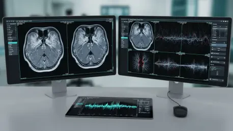

For more than a century, neurologists have known that the cognitive decay associated with multiple sclerosis stems from damage to the brain’s gray matter, yet these vital areas remained stubbornly invisible on conventional MRI scans until the arrival of sophisticated computational models. The AI-driven MS neuroimaging represents a significant advancement in the medical imaging and neurology sectors. This review explores the evolution of the technology, its key features, performance metrics, and the impact it has had on various applications. The purpose of this review is to provide a thorough understanding of the technology, its current capabilities, and its potential development. By applying advanced computational processing to standard magnetic resonance imaging scans, software-based analysis is now capable of identifying pathology that was previously detectable only through postmortem histopathology.
Foundations of AI-Enhanced Neuroimaging in Multiple Sclerosis
This technology introduces a paradigm shift in how clinicians approach the diagnosis and monitoring of Multiple Sclerosis. Traditionally, magnetic resonance imaging served as the gold standard but suffered from a “gray matter gap,” where lesions in the cerebral cortex remained largely invisible to the human eye. The emergence of AI-driven models has bridged this gap by shifting the focus from hardware-strength limitations to sophisticated software-based analysis. Rather than relying on more powerful magnets, which are expensive and often inaccessible, these models extract more value from existing data.
The evolution of these tools places AI at the center of the modern neurological landscape, transforming “invisible” disease indicators into quantifiable clinical data. This shift is critical because cortical damage is the most accurate predictor of long-term physical disability and cognitive decline. By providing a clear view of these areas, the technology allows for a more holistic understanding of the disease, moving beyond the traditional focus on white matter inflammation. This foundational change ensures that clinicians no longer overlook the silent drivers of the condition.
Advanced Architectural Components of AI-Driven Imaging
Multimodal Cortical Lesion Enhancement (MMCLE)
MMCLE serves as the primary technical engine for identifying gray matter damage. This component functions by synthesizing information across multiple MRI sequences, rather than relying on a single image contrast. By analyzing how tissue behaves across different scans, the system can highlight abnormalities that lack sufficient contrast on conventional MRI. This multimodal approach allows the AI to “look between” the data points, identifying minor discrepancies in signal intensity that suggest tissue degradation.
The performance of MMCLE is particularly noteworthy because it provides a high-resolution method for detecting thousands of previously overlooked lesions per patient group. It effectively increases the signal-to-noise ratio in regions of the brain where the contrast between healthy and diseased tissue is notoriously slim. This architectural component is what differentiates modern AI solutions from traditional automated segmentation tools, which often fail when faced with the subtle textures of the cerebral cortex.
Generative AI and Legacy Data Synthesis
A critical feature of this technology is its ability to utilize “legacy” or existing MRI data. Instead of requiring new, high-cost imaging hardware, generative AI models can process retrospective scans from past clinical trials or routine hospital visits. This technical aspect allows for the reconstruction of a patient’s neurological health profile using standard clinical hardware. The significance of this component lies in its ability to offer high-resolution insights without the need for expensive equipment upgrades, making advanced diagnostics more accessible to a wider range of medical facilities.
By synthesizing older data, these models can create longitudinal views of disease progression that were previously impossible to generate. This capability is essential for understanding how a patient has reached their current state and for evaluating the long-term effectiveness of various therapies. The ability to pull meaningful insights from lower-resolution legacy scans ensures that the benefits of AI are not restricted to top-tier research institutions, democratizing advanced neurology.
Current Trends and Innovations in Neural Pattern Recognition
The field is currently moving toward an “intelligence-first” model, where software innovation takes precedence over magnet strength in MRI machines. There is a growing industry shift toward monitoring “silent progression,” the cognitive and physical decline that occurs even when white matter appears stable. Recent developments emphasize the use of AI to standardize the detection of cortical lesions, which were only recently added to formal diagnostic criteria. This trend reflects a broader move toward deep-learning models that can interpret the subtle variations in tissue texture that define gray matter pathology.
Moreover, the industry is seeing a convergence of neuroimaging with other biomarkers. AI models are increasingly being trained to correlate imaging findings with genomic data and fluid biomarkers, such as neurofilament light chain levels. This holistic approach to pattern recognition allows for a more comprehensive assessment of the patient’s status. The innovation lies in the AI’s ability to find correlations across these diverse data sets that would be impossible for a human researcher to identify manually.
Real-World Applications in Clinical and Research Settings
AI-driven neuroimaging is being deployed extensively in pharmaceutical research, notably in large-scale Phase III clinical trials such as those for Ocrelizumab. In these settings, the technology provides a more accurate metric for drug efficacy, specifically regarding how well a treatment protects the brain’s gray matter. The ability to detect thousands of cortical lesions across a trial population gives researchers a much more sensitive tool for measuring neuroprotection, which is a primary goal of next-generation MS therapies.
In clinical practice, these models are used to provide patients with a clearer prognosis by revealing the “hidden” drivers of long-term disability. This is particularly relevant in the neurology sector, where early intervention in cortical damage can significantly alter a patient’s quality of life and cognitive outcomes. By visualizing the full extent of the disease, physicians can make more informed decisions about treatment escalations or changes in therapeutic strategy, ensuring that the management plan matches the actual severity of the pathology.
Technical Hurdles and Market Obstacles
Despite its success, the technology faces challenges regarding the variability of MRI hardware across different medical facilities. Technical hurdles include ensuring that AI models remain accurate when processing scans from different manufacturers or older machines with varying magnetic field strengths. The “black box” nature of some deep-learning models also remains a concern for clinicians who require explainable results to make treatment decisions. Ensuring that an AI’s findings can be verified and understood by a radiologist is a key requirement for broader adoption.
Additionally, there are regulatory and integration obstacles to overcome before these AI tools can be seamlessly embedded into everyday clinical workflows. Standardizing the output of these models so they can be easily interpreted across different hospital systems is a significant logistical task. Ongoing development efforts are focused on streamlining image-processing techniques to make them faster and more “plug-and-play” for radiologists who may not have specialized computational expertise, reducing the friction of implementing these advanced tools.
Future Outlook and Long-Term Technological Trajectory
The future of MS neuroimaging is heading toward a fully personalized medicine approach. Potential breakthroughs include AI models that can predict the specific trajectory of a patient’s disability years in advance by analyzing early cortical changes. This predictive power would allow for the implementation of preventative strategies long before physical symptoms manifest. As these tools become more refined, they are expected to lead to the development of a new generation of therapeutics specifically designed to preserve gray matter rather than just reducing inflammation.
Furthermore, the long-term impact on society will likely be a reduction in the burden of MS-related disability and a significant improvement in cognitive health management for millions of patients. The trajectory of this technology suggests a move toward real-time monitoring, where AI-integrated wearable devices and frequent, low-cost imaging provide a continuous stream of data. This would transform MS management from a series of reactive snapshots into a proactive, data-driven journey of preservation.
Final Assessment of AI Integration in Neurology
The integration of AI into MS neuroimaging marked a monumental advance in the ability to visualize the full extent of the disease. By turning previously invisible pathology into actionable data, this technology addressed a century-old limitation in neurology. The performance in both retrospective research and prospective clinical monitoring demonstrated that software intelligence could effectively compensate for hardware constraints. The transition to AI-integrated diagnostics necessitated a total overhaul of imaging protocols and demanded that pharmaceutical companies re-evaluate their long-term clinical endpoints.
Looking ahead, the clinical community must focus on the universal standardization of AI outputs to ensure equitable access to these diagnostic breakthroughs across different geographic regions. The next logical step involved the integration of these imaging tools into routine electronic health records, allowing for automated alerts when subtle cortical changes were detected. As the healthcare industry continued to adopt these models, the focus shifted from simple lesion detection toward complex predictive analytics, ensuring that no aspect of MS-related damage remained hidden from clinical view.