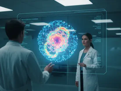

The conventional diagnostic approach of treating a tumor as a uniform mass of malignant cells has reached a functional ceiling in oncology, necessitating a move toward high-resolution architectural mapping. The Spatial Transcriptomic Tumor Atlas represents a pivotal shift in this landscape, moving beyond the limitations of bulk RNA sequencing which typically averages genetic signals across thousands of cells. By preserving the geographical context of gene expression, this technology allows clinicians to see not just which mutations are present, but exactly where they are located within the complex tumor microenvironment.
This evolution is fundamentally driven by the failure of traditional molecular subtyping to predict treatment outcomes consistently. While older methods classified tumors into static categories, the spatial approach recognizes the tumor as a dynamic ecosystem. It effectively bridges the gap between histopathology, which looks at structure, and genomics, which looks at instructions. By integrating these disciplines, the atlas provides a granular view of the “cellular conversations” that drive cancer progression and therapeutic resistance.
Evolution and Core Principles of Spatial Transcriptomics in Oncology
The emergence of spatial transcriptomics marks a departure from the “smoothie” approach of bulk sequencing, where tissue is homogenized and spatial context is lost. In contrast, this technology functions like a high-definition map, pinning transcriptomic data to specific coordinates on a tissue slide. This allows for the identification of rare cell populations and localized signaling pathways that were previously obscured by the dominant signals of more abundant cells.
In the broader technological landscape, this methodology addresses the clinical “black box” of tumor heterogeneity. Traditional methods often fail because they treat a biopsy as representative of the entire malignancy. This technology overcomes such limitations by visualizing how different regions of a single tumor may possess vastly different molecular profiles, explaining why a patient might respond to treatment in one area while progressing in another.
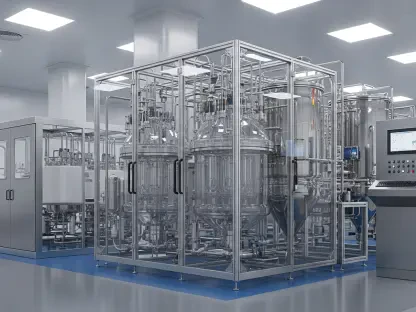
Technical Framework and Data Integration Strategies
Integrated Multi-Omic Data Acquisition
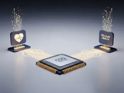
The technical superiority of this atlas stems from its multi-omic synthesis, combining spatial transcriptomics with whole-exome sequencing and single-cell RNA sequencing. This integration is crucial because transcriptomics alone only reveals what a cell is doing, whereas genomics reveals the underlying mutations driving those actions. By layering these data sets, researchers can correlate specific genetic aberrations with their physical manifestations in the tumor architecture.
This multi-dimensional view is unique because it synchronizes disparate data types into a unified coordinate system. While competitors might focus on a single modality, this implementation uses single-cell data to “deconvolve” the spatial signals, providing a more accurate estimation of cell-type abundance at every location. This ensures that the biological insights are not just observational but are grounded in the mechanical reality of the tumor’s genetic makeup.
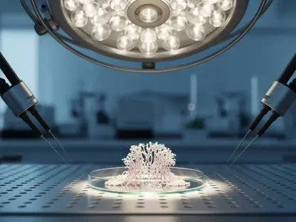
High-Resolution Mapping and Validation
Creating a reliable spatial atlas requires rigorous validation across large, independent cohorts to ensure that observed patterns are not artifacts of a single sample. The use of pretreatment tumor samples is essential here, as it provides a baseline of the tumor’s native state before therapeutic intervention alters the landscape. This baseline is critical for identifying the primary drivers of the disease and potential “escape routes” the cancer might take.
The validation process involves comparing spatial patterns across thousands of tumors, which transforms a localized observation into a clinically relevant biomarker. This high-resolution mapping ensures that the spatial signatures identified are robust and reproducible. Such a comprehensive approach is what separates this technology from experimental academic exercises, positioning it as a viable tool for prospective clinical decision-making.
Emerging Trends in Tumor Subtyping and Cellular Differentiation
The field is currently moving away from the rigid binary classification of tumors, such as the luminal versus basal distinction. Instead, the spatial atlas reveals a continuous differentiation axis, suggesting that cancer cells exist in a spectrum of states rather than fixed categories. This nuance is critical for precision medicine, as it acknowledges that a single tumor can harbor multiple identities simultaneously, often transitioning between them based on local environmental cues.
This discovery of coexisting cell states is fundamentally changing research priorities within the industry. Scientists are no longer looking for a “one-size-fits-all” marker but are instead focusing on how to manage a shifting cellular landscape. This trend toward “plasticity awareness” suggests that future therapies will need to be as flexible as the tumors they are designed to treat, targeting the transitions between states rather than just the states themselves.
Real-World Applications in Precision Medicine
Targeted Strategies for Luminal Tumor Cores
Within the spatial architecture, luminal-like regions are often found in the “immune-cold” cores of the tumor, where they are shielded from the body’s natural defenses. The atlas has identified specific markers like NECTIN4 and FGFR3 as being highly enriched in these protected areas. This provides a clear therapeutic target for antibody-drug conjugates, which can penetrate these dense cores to deliver localized treatment where traditional immunotherapy might fail.
Identifying these vulnerabilities matters because it allows for the deployment of “surgical” molecular strikes. Instead of broad-spectrum chemotherapy, clinicians can use the spatial map to determine if a patient has a dominant luminal core that would be sensitive to targeted inhibitors. This implementation is unique in its ability to predict resistance by showing where the target is absent, even if a bulk test says the marker is present overall.
Immunotherapy and Management of Basal Margins
In contrast to the cold cores, the invasive margins of many tumors often exhibit a basal-like, “inflamed” phenotype. These regions are highly aggressive but also more accessible to the immune system, making them prime candidates for checkpoint inhibitors. The spatial atlas helps in visualizing this “battlefront,” allowing doctors to see how immune cells are interacting with the tumor’s edge and whether those immune cells are being suppressed.
This spatial insight is vital for managing aggressive disease, as it explains why some “inflamed” tumors still resist treatment. It might be that the immune cells are present but physically excluded from the tumor’s interior by a dense fibrous barrier. By mapping these physical obstacles, the atlas provides a rationale for combination therapies that can break down these barriers or enhance immune infiltration at the margins.
Technical Hurdles and Clinical Implementation Challenges
Despite its potential, the technology faces significant hurdles, primarily the need for even larger prospective clinical cohorts to prove its predictive value. Analyzing the tumor landscape post-treatment also remains difficult, as the architecture is often disrupted by cell death and scarring, making it hard to track how the cancer evolved in response to the drug. This “post-treatment void” is a major area of ongoing development.
Furthermore, the complexity of analyzing such massive data sets requires specialized bioinformatics expertise that is not yet available in every clinical setting. There is also the persistent challenge of “sampling bias” in single-site biopsies. While the atlas provides a better map, it still relies on the quality of the initial tissue sample. Ongoing efforts to create “spatially informed biomarkers” aim to mitigate this by identifying liquid biopsy signatures that reflect these complex spatial distributions.
Future Outlook and Longitudinal Research
The future of this technology lies in its transition from a static map to a longitudinal tracking system. By observing how therapies reshape tumor architecture over time, researchers can begin to decode the “cellular language” of resistance. This would allow for the development of adaptive treatment plans that change in real-time as the tumor’s spatial organization shifts, preventing the emergence of resistant clones before they can take over.
The long-term impact on precision medicine will be a move toward truly individualized oncology. As the cost of spatial sequencing continues to decrease, it is likely that every major cancer center will utilize these atlases to guide complex cases. This will lead to a more sophisticated understanding of the tumor microenvironment, where the physical location of a cell is considered just as important as its genetic sequence in determining the best course of action.
Summary and Final Assessment of the Spatial Atlas
The development of the spatial transcriptomic tumor atlas successfully dismantled the traditional binary subtyping framework by proving that luminal and basal states often coexist within a single malignancy. The project revealed that the physical organization of these states was not random but followed a predictable differentiation axis that correlated with therapeutic sensitivity. By mapping markers like NECTIN4 and FGFR3 to specific geographic regions, the research established a foundation for more precise treatment sequencing.
The analysis underscored the necessity of spatial awareness in oncology, as it explained the frequent failures of single-biopsy diagnostics. It was concluded that future clinical trials must incorporate spatial biomarkers to accurately categorize patient response and identify localized resistance mechanisms. Moving forward, the integration of this technology into routine clinical practice promised to transform bladder cancer treatment from a series of educated guesses into a data-driven strategy. This shift significantly advanced the industry’s ability to manage tumor heterogeneity and improved the outlook for individualized patient care.