
Surgeons have long struggled with the intricate biological demands of repairing massive skeletal defects where traditional bone grafts often fail due to poor integration or limited availability of donor tissue. In the current landscape of regenerative medicine, the pursuit of a biocompatible scaffold that mimics the natural extracellular matrix has led to a revolutionary development in DNA-based hydrogels. These materials are not merely passive fillers; they serve as dynamic environments that can be programmed to release growth factors at precise intervals. By utilizing the inherent programmability of nucleic acids, bioengineers have created a three-dimensional architecture that guides cellular migration and differentiation with unprecedented accuracy. This breakthrough addresses the critical gap in treating non-union fractures and complex trauma where the body’s natural healing processes are insufficient. As clinical trials progress through 2026 and into 2027, the potential to replace invasive metal hardware with bio-absorbable, self-assembling structures is becoming a tangible reality for patients worldwide.
The Mechanics of Molecular Scaffolding
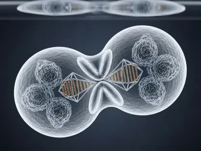
The structural integrity of these new hydrogels relies on the precise self-assembly of synthetic DNA strands that are engineered to interlock into a robust yet porous matrix. Unlike traditional polymer-based scaffolds, these DNA-based systems use specific nucleotide sequences to dictate the physical properties of the gel, such as its stiffness and degradation rate. This level of control allows for the customization of the scaffold to match the mechanical requirements of different bone types, from the dense cortical bone of the femur to the more porous cancellous bone of the vertebrae. Furthermore, the hydrogel acts as a reservoir for bioactive molecules, such as Bone Morphogenetic Protein-2, which is released gradually as the DNA scaffold is broken down by the body’s natural enzymes. This synchronized degradation ensures that as the synthetic material disappears, it is replaced by freshly mineralized bone tissue. This approach minimizes the risk of chronic inflammation often associated with permanent synthetic implants.
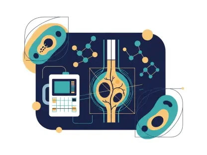
Beyond physical support, the DNA hydrogel facilitates a sophisticated communication network between the scaffold and the surrounding biological environment. Endogenous stem cells are recruited to the site of injury through chemotactic signals embedded within the DNA strands, effectively turning the injury site into a localized bio-factory for bone production. The high water content and biocompatibility of the hydrogel prevent the immune system from identifying the scaffold as a foreign threat, which significantly reduces the incidence of graft rejection. Researchers have observed that vascularization—the formation of new blood vessels—is greatly accelerated within these DNA frameworks compared to ceramic or metallic alternatives. This is a crucial factor in bone healing, as a steady supply of oxygen and nutrients is necessary to sustain the high metabolic demands of osteoblasts during the mineralization phase. By creating a hospitable microenvironment that actively participates in the healing process, these hydrogels set a new standard for reconstructive surgery and recovery protocols.
Clinical Implications: Transitioning From Laboratory to Operating Room
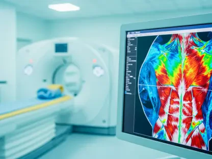
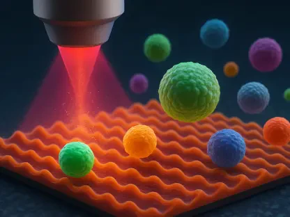
The shift toward utilizing DNA-based materials in clinical settings represents a significant departure from the “one-size-fits-all” approach of traditional orthopedic implants. Surgeons are now looking at personalized treatment plans where the DNA hydrogel can be 3D-printed or injected directly into a void, conforming perfectly to the unique geometry of a patient’s injury. This precision reduces the surgical time and the complexity of the procedure, as there is less need for manual shaping of bone grafts or the installation of extensive fixation hardware. In recent pilot studies, patients treated with these programmable hydrogels showed a forty percent increase in bone density within the first six months compared to those receiving standard care. The implications for aging populations, particularly those suffering from osteoporosis or related degenerative conditions, are profound. By providing a scaffold that effectively “tricks” the body into aggressive self-repair, medical professionals can achieve successful outcomes in cases previously deemed untreatable or high-risk for permanent disability.
The successful implementation of DNA hydrogels in complex bone regeneration established a foundational shift in how traumatic injuries and degenerative diseases were approached. Medical institutions prioritized the integration of bio-printing facilities within surgical departments to allow for the real-time customization of these biological scaffolds. To ensure sustained success, healthcare providers invested in specialized training for orthopedic surgeons to master the delivery and monitoring of these bioactive materials. The focus shifted toward long-term data collection to refine the DNA sequences used for specific metabolic profiles, ensuring that regenerative therapies remained optimized for individual patient needs. Future considerations included the expansion of this technology to repair cartilage and ligament tissues, which often presented similar regenerative challenges. By adopting these advanced bio-materials, the medical community successfully moved toward a paradigm where permanent metal implants became a secondary option, favoring instead the total biological restoration of the skeletal system for improved long-term mobility.