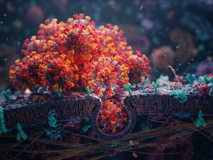

Small-scale nanoparticles are uniquely capable of penetrating the dense architecture of biofilms to disrupt the quorum sensing communication that bacteria use to maintain their survival. This revelation comes at a time when the global medical community is facing a significant crossroads, as the standard arsenal of antibiotics continues to lose ground against increasingly sophisticated and resilient superbugs. For decades, the pharmaceutical model relied heavily on the discovery of new small-molecule inhibitors that target specific bacterial pathways, but the inherent adaptability of microbes has rendered many of these once-miraculous drugs ineffective in modern clinical settings. The scale of this crisis is profound, with current health projections indicating that by the middle of the century, resistant infections could claim millions of lives annually and cause trillions of dollars in economic disruption. Consequently, researchers are pivoting toward inorganic chemistry and nanotechnology, where metallic nanoparticles (MNPs) offer a revolutionary paradigm shift. Unlike traditional antibiotics, these particles use a multifaceted attack strategy, overwhelming bacteria through multiple simultaneous mechanisms that make the development of resistance far less likely for the organism.
Methods for Creating Nanoscale Antimicrobials
Conventional and Sustainable Synthesis Techniques
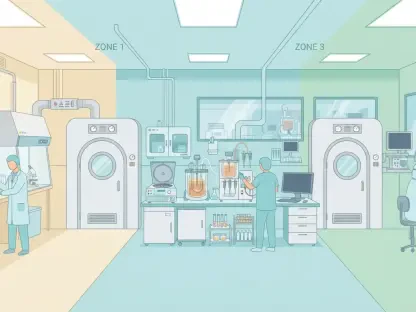
Chemical reduction remains the primary industry standard for creating these nanoscale tools, a process that relies on the precise transformation of metal salts into metallic clusters through controlled chemical reactions. This method typically involves using a reducing agent, such as sodium borohydride or ascorbic acid, to manipulate the oxidation states of metal salts like silver nitrate or copper sulfate until stable nanoparticles begin to form. To maintain the structural integrity and specific size of these particles, researchers must introduce capping agents or stabilizers that coat the surface and prevent the particles from aggregating into larger, less effective masses. While this approach offers unparalleled scalability and precision for manufacturing silver, gold, and copper agents, it is not without its complications. The reliance on potentially toxic chemical precursors often leaves residual impurities that can compromise the biocompatibility of the final therapeutic product, necessitating extensive purification steps. As the demand for clinical-grade nanomaterials grows, the environmental and safety profile of these chemical routes is coming under increasing scrutiny, forcing scientists to balance manufacturing efficiency with the need for non-toxic materials.
In response to the limitations and environmental footprint of chemical reduction, green synthesis has emerged as a transformative and eco-friendly alternative that leverages the natural chemistry of the biosphere. By utilizing extracts from plants, fungi, and bacteria as both reducing and stabilizing agents, scientists have successfully produced nanoparticles that are inherently more compatible with biological systems. For example, recent studies have demonstrated that simple biological precursors like onion broth or fungal filtrates can facilitate the creation of silver nanoparticles that are highly active against common pathogens such as E. coli. These biological pathways are particularly attractive because the resulting particles are often pre-coated with natural phytochemicals and proteins that enhance their stability without the need for synthetic additives. However, it is essential to recognize that a natural origin does not automatically guarantee that a substance is safe for systemic human use. The scientific community is currently working to standardize these biological processes, as the inherent variability in plant and fungal extracts can lead to inconsistencies in particle size and potency that must be addressed before mass clinical adoption becomes a reality.
Specialized Production: For Enhanced Performance
Beyond the standard chemical and biological routes, advanced physical methods such as microwave-assisted synthesis have provided a way to create highly uniform particles with remarkable speed and efficiency. This technique uses electromagnetic radiation to provide rapid, localized heating, which ensures a more even distribution of energy throughout the reaction mixture than conventional heating methods could ever achieve. The result is a batch of nanoparticles with extremely narrow size distributions, which is critical for ensuring consistent antimicrobial performance in medical applications. Similarly, the hydrothermal method has gained traction for its ability to produce highly crystalline structures by subjecting metal precursors to intense pressure and elevated temperatures in a sealed environment. These specialized physical approaches allow for the creation of hybrid materials and complex metal oxides that possess superior structural integrity. By fine-tuning the synthesis parameters, researchers can effectively control the exact physical characteristics of the nanoparticle to maximize its ability to interact with and destroy resistant pathogens.
This high level of control is particularly evident in the development of sophisticated materials like cobalt ferrite and magnesium oxide nanoparticles, which have shown impressive results in inhibiting stubborn Gram-positive bacteria like Staphylococcus aureus. The sol-gel process is another technique highlighted for its utility in creating these metal oxides, allowing for the formation of particles that can be easily integrated into medical coatings or wound dressings. These advanced production methods are not merely about creating smaller particles; they are about engineering functionalized surfaces that can trigger specific biological responses. For instance, hydrothermal synthesis can be used to create nanoparticles with sharp, needle-like geometries that physically puncture bacterial membranes upon contact. As the field moves toward more personalized and targeted antimicrobial therapies, the ability to manufacture these diverse and highly specialized nanostructures will be the deciding factor in how effectively we can combat specific classes of drug-resistant bacteria in a clinical setting.
How Nanoparticles Destroy Resistant Bacteria
Physical Damage: And Oxidative Stress
The primary way metallic nanoparticles eliminate bacteria is through a direct and devastating physical interaction with the cell wall that traditional antibiotics simply cannot replicate. Most bacterial species possess a negatively charged outer surface, which acts as a natural magnet for positively charged metallic nanoparticles introduced into the environment. Upon making contact, these particles can warp the structural integrity of the cell membrane, leading to the immediate leakage of vital internal components such as ions and proteins. In many cases, the physical force exerted by the nanoparticles is so intense that it causes the membrane to rupture entirely, killing the microbe before it has a chance to mount a biological defense. Some materials, such as magnesium oxide, have proven to be so effective that they can neutralize a pathogen simply by adhering to its outer surface, demonstrating that physical contact alone is often sufficient to end a bacterial infection without the particle ever needing to enter the cytoplasm.
Another formidable weapon in the nanoparticle arsenal is the induction of overwhelming oxidative stress through the generation of reactive oxygen species (ROS). When metallic nanoparticles interact with the cellular environment, they can catalyze the formation of highly volatile radicals, including superoxide, hydrogen peroxide, and hydroxyl radicals. When the production of these radicals exceeds the bacteria’s natural antioxidant defenses, the results are catastrophic for the microbe’s survival. These radicals initiate lipid peroxidation in the cell membranes, disrupt the delicate process of protein folding, and cause irreversible breaks in the bacterial DNA. This “oxidative blast” is particularly potent because it attacks multiple cellular targets simultaneously, making it virtually impossible for a bacterium to develop resistance through a single genetic mutation. Different metals are known to produce different types of ROS; for instance, silver and copper generate a broad spectrum of destructive radicals, whereas zinc oxide focuses its energy on specific cellular processes to achieve its antimicrobial effect.
Internal Interference: And Biofilm Eradication
Once a nanoparticle or its released ions successfully penetrate the interior of a bacterial cell, they begin to systematically dismantle the organism’s internal life-support systems. These metal ions have a high affinity for functional groups such as thiols, phosphates, and amines, which are the building blocks of essential enzymes and proteins. By binding to these sites, the metallic agents deactivate the enzymes required for metabolism and disrupt the ribosome’s ability to synthesize new proteins, effectively halting the bacteria’s growth and replication cycle. Gold nanoparticles, in particular, have been observed to collapse the membrane potential of pathogens like E. coli by specifically blocking the activity of ATPase, the enzyme responsible for energy production. This targeted internal interference ensures that even if a bacterium manages to survive the initial physical damage to its outer shell, its core metabolic machinery is systematically shut down, leading to an inevitable and total cellular collapse.
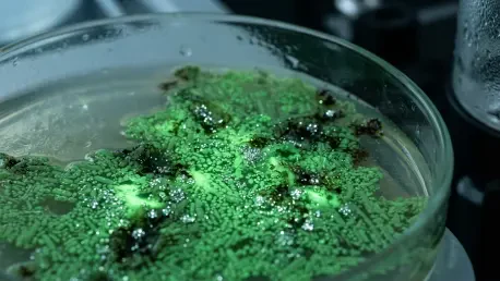
In addition to killing individual bacterial cells, metallic nanoparticles show incredible promise in tackling biofilms, which are slimy, protective colonies that are notoriously difficult to treat with standard medicine. Biofilms act as a physical shield, making the bacteria within them up to a thousand times more resistant to conventional antibiotics than free-floating cells. Because of their incredibly small size, nanoparticles are able to slip through the dense molecular architecture of these biofilms to reach the pathogens hidden deep inside. Once they have gained entry, they release ions that disrupt the “quorum sensing” communication that bacteria use to coordinate the maintenance and growth of the biofilm matrix. Research has highlighted that when metallic nanoparticles are combined with natural bioactive compounds like curcumin, their ability to dissolve these protective shields is significantly enhanced. This synergistic approach not only breaks down the physical barrier of the biofilm but also leaves the underlying bacteria exposed and vulnerable to the antimicrobial action of the nanoparticles.
Factors Influencing Efficacy and Safety
Physical Traits: And Biological Barriers
The overall effectiveness of a metallic nanoparticle is heavily dictated by its specific physical dimensions and its geometric shape, both of which determine how it interacts with biological matter. Generally, smaller particles perform significantly better in antimicrobial tests because they possess a much higher surface-area-to-volume ratio, which allows for more extensive interaction with bacterial membranes and a faster release of toxic metal ions. However, the size of the particle also influences how it is perceived by human cells, with the smallest particles sometimes being internalized by healthy tissue more easily than their larger counterparts. The geometry of the particle—whether it is shaped like a sphere, a rod, or a triangle—also plays a critical role in its ability to penetrate bacterial defenses. For example, triangular silver nanoparticles have been shown to have a higher degree of reactivity and a greater ability to puncture cell walls than spherical ones, suggesting that “sharp” nanostructures may be inherently more lethal to pathogens.
Furthermore, the specific type of bacteria being targeted is a major factor in determining how a nanoparticle should be engineered for maximum success. Gram-negative bacteria, such as E. coli, possess an additional outer membrane that acts as a sophisticated barrier against many foreign substances, often making them more resistant to treatment than Gram-positive strains. Yet, certain nanomaterials like zinc oxide have shown a surprising level of toxicity toward Gram-positive bacteria, indicating that the interaction between the nanoparticle surface and the bacterial cell wall is highly strain-specific. This variability highlights the need for a more nuanced approach to nanomedicine, where particles are tailored to the unique biological signatures of the infection they are intended to treat. Understanding these physical and biological determinants is essential for moving beyond a “one-size-fits-all” approach and toward a future where nanotechnology can be used to precisely target and eliminate specific resistant pathogens without causing collateral damage to the host.
Translational Challenges: The Path to Clinical Safety
Despite the undeniable success of metallic nanoparticles in laboratory settings, the transition to real-world clinical use is blocked by several significant safety concerns and biological hurdles. One of the most prominent issues is the “protein corona” effect, which occurs the moment a nanoparticle enters the human bloodstream or wound fluid. In these environments, various proteins immediately coat the surface of the nanoparticle, forming a thick layer that can completely change the particle’s surface charge and biological identity. This coating can mask the nanoparticle’s intended antimicrobial powers or, in some cases, trigger an unintended immune response from the host. Additionally, the tendency of nanoparticles to aggregate in the presence of natural salts found in the human body can reduce their efficacy and change how they are distributed throughout the patient’s organs. These complex interactions within the biological environment mean that a particle that performs perfectly in a test tube might behave in an entirely unpredictable and potentially dangerous manner once injected into a living system.
The long-term safety and eventual disposal of metallic agents within the human body also represent a major regulatory hurdle for the pharmaceutical industry. There is a very narrow window between the concentration of metal required to kill a pathogen and the concentration that becomes toxic to the host’s own mitochondria and DNA. Concerns regarding the accumulation of metals like silver, copper, or zinc in vital organs such as the liver and kidneys have not yet been fully addressed through long-term clinical studies. Furthermore, the global medical community lacks a standardized set of international protocols for testing the antimicrobial activity of nanomaterials, leading to wildly inconsistent results across different research facilities. There is also the emerging threat of “nanotolerance,” where bacteria may eventually develop specialized efflux pumps to remove metal ions from their systems. Addressing these toxicological and regulatory challenges is the next necessary step in ensuring that metallic nanoparticles can be safely integrated into the modern medical workflow as a reliable solution for the antibiotic crisis.
Future Strategies for Nanomaterial Integration
The exploration of metallic nanoparticles provided a clear view into a new era of infection control where inorganic materials served as the frontline defense against resistant superbugs. Researchers established that the multi-targeted mechanisms of these particles offered a significant advantage over the narrow focus of traditional antibiotics, effectively raising the bar for bacterial survival. The study of various synthesis methods, from chemical reduction to green biological routes, highlighted the importance of balancing manufacturing efficiency with the rigorous safety standards required for human health. It was concluded that the success of these materials depended not just on their ability to kill bacteria in a laboratory, but on their behavior within the complex biological landscape of the human body.
Moving forward, the focus must shift toward the development of standardized toxicological frameworks and the implementation of long-term animal models to track the movement and accumulation of metals in living tissue. Scientists suggested that the integration of nanoparticles into existing medical infrastructure, such as smart wound dressings and antimicrobial coatings for surgical implants, represented the most immediate and viable path for clinical adoption. The findings emphasized that while metallic nanoparticles were a powerful tool, they required precise engineering to overcome the challenges of the protein corona and potential bacterial tolerance. By prioritizing the creation of biocompatible, hybrid materials and fostering international cooperation on regulatory standards, the medical community took the necessary steps to turn the promise of nanotechnology into a practical reality for patients worldwide.