
For patients living with a chronic Achilles rupture that has resisted months of therapy and bracing, every step can feel like a wager against pain, weakness, instability, and the fear that a compromised tendon will fail again during even the simplest tasks. Traditional reconstruction can fix the gap, yet the path to that outcome often runs through complex surgery, wound risks, and weeks of guarded motion. A different proposition has quietly taken shape: a single injection of a patient’s own bone marrow–derived mesenchymal stem cells, expanded in a cleanroom and placed under imaging guidance into the rupture zone. Rather than patching with grafts or moving another tendon, this strategy aims to coax the injured ends to knit by rebuilding aligned fibers. Early clinical observations from a small case series at the Institute of Tissue Regenerative Therapy offered a window into what such a biologic repair could look like if carefully selected patients were treated and mobilized early.
Who Was Treated and How the Therapy Was Delivered
The cohort consisted of six adult men with chronic Achilles ruptures that had not improved with prior care, a group often facing reconstruction or tendon transfer. Investigators harvested bone marrow, isolated mesenchymal stem cells, and cultured them for roughly 22 days in a controlled cleanroom to reach a target dose of about 20 million viable cells per patient. Because the cells were autologous, donor matching and most immune compatibility issues were avoided, simplifying logistics at the bedside. Under image guidance, clinicians injected the expanded cells directly into the rupture gap, aiming to populate the defect and influence the healing milieu. Patients stood and walked immediately after the procedure and advanced through supervised strengthening without casts or boots. Daily routines resumed the next day, with a careful ramp to recreational sport by about four months, reflecting confidence in the tendon’s evolving load tolerance.
What Imaging and Clinical Outcomes Showed
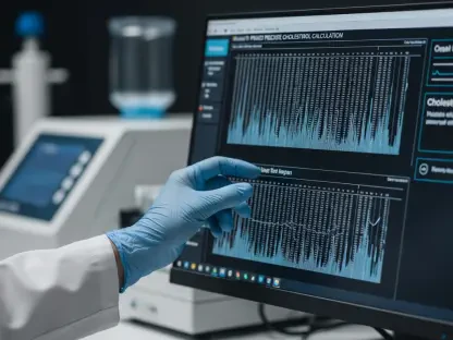
Magnetic resonance imaging supplied a structural narrative that matched the clinical course, documenting progressive filling of the gap and the reappearance of aligned tendon fibers across the original defect. Reported median regeneration reached 96.3% at 12 months and 100% by 24 months in all six cases, with no calcifications or aberrant deposits noted during follow-up. Interestingly, symptoms improved faster than images transformed: patients described meaningful pain relief by three months, and by the final visit, pain scores for both daily activities and sports had fallen to zero. Function tracked the same arc. The Achilles score rose sharply, with a median gain of 86.5 points, signaling substantial recovery in tasks that strain the tendon. The early permission to bear weight did not precipitate setbacks; instead, it seemed to coincide with steady structural maturation, though causality remained uncertain without a controlled comparator.
Why Mesenchymal Stem Cells Might Help
The biological rationale focused less on the cells becoming tendon and more on their capacity to choreograph the local repair process. Mesenchymal stem cells are known to secrete a suite of paracrine factors that can dial down inflammation, spur angiogenesis, and direct resident tenocytes to lay down organized collagen rather than scar. In a degenerative, chronic rupture bed—where disarray and poor vascularity stifle healing—such signals may reset the microenvironment toward alignment and continuity. Reviews referenced by the treating group have argued that this modulation can produce sturdier, more elastic tissue than passive scarring, which often widens under load. Yet key unknowns remained: how long the injected cells persisted in human tendons, which signals drove durable remodeling, and whether the roughly 20 million–cell dose was optimal. Delivery nuances, including needle trajectory and dispersion within the gap, also awaited systematic testing.
How This Approach Compares With Standard Surgery
Chronic Achilles repairs challenge even experienced surgeons because tendon ends retract and interpose scar, often necessitating V‑Y lengthening, turndown flaps, or flexor hallucis longus transfer. A frequently cited synthesis placed complication rates around 11.4% for such reconstructions, with wound problems leading the list, a predictable concern given limited soft‑tissue coverage above the tendon. By contrast, a validated, needle‑based strategy could, in theory, trim morbidity, avoid new incisions, and enable immediate mobilization that preserves calf conditioning. The ITRT series hinted at that possibility, coupling rapid pain decline with uninterrupted daily activity and no immobilization. However, surgical reconstruction has decades of outcomes across demographics and athletic tiers, while cell injections remained nascent. Only head‑to‑head, controlled studies could determine equivalence or superiority for specific patient profiles, gap sizes, and timelines from injury, and define trade‑offs that matter to patients.
Strength of Evidence and Safety Signals to Date
The case series fit within a growing movement in sports medicine toward guided biologic repair, complementing mechanical strategies rather than replacing them outright. Prior work from the same group in the patellar tendon suggested bone marrow cell therapy could outperform a blood‑based injection in structural gains, hinting that true cell‑based approaches might exceed simpler biologics like platelet‑rich plasma in certain contexts. Importantly, the Achilles report tied symptom relief to MRI‑verified fiber continuity, aligning with an emerging consensus that both imaging and validated function scores are needed to claim genuine regeneration. Safety signals were favorable: over at least two years, no serious adverse events or re‑ruptures were recorded, and no ectopic calcification appeared. Even so, the tiny, homogeneous cohort—six Caucasian men at a single site—meant rare harms could have been missed, and external validity remained limited pending broader sampling.
What Must Be Tested Before Broader Adoption
A concrete roadmap had emerged. Randomized or rigorously matched trials should compare this injection against reconstruction, nonoperative protocols, and alternative biologics, enrolling women and men across age ranges and activity demands, from desk workers to high‑impact athletes. Protocols need factorial testing of dose, culture duration, delivery technique, and staged rehabilitation, with standardized MRI, ultrasound where appropriate, validated function scores, and activity‑specific endpoints tracked for multiple years from 2026 to 2028 and beyond. Manufacturing quality, cost transparency, and regulatory compliance for expanded autologous cells must be integrated early, or scalability would falter. Clinicians could have prioritized shared decision‑making that framed this option as experimental, reserved for select chronic ruptures with informed consent. Health systems might have prepared registries to capture rare events. By taking these steps, the field positioned itself to decide whether a needle could consistently match a scalpel for this stubborn injury.