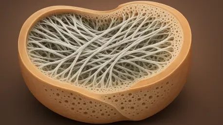

Researchers at Birjand University of Medical Sciences are targeting the unique mechanical requirements of the knee by leveraging the dense native collagen found in camels. This specific focus on camel-derived tissue addresses one of the most persistent challenges in orthopedic medicine: the repair of the human meniscus. As a crescent-shaped wedge of fibrocartilage, the meniscus is essential for shock absorption and joint stabilization, yet its biological nature makes it incredibly difficult to heal. The inner two-thirds of the structure are avascular, meaning they lack the blood supply necessary for natural regeneration. Consequently, injuries caused by sports trauma or the degeneration of aging often result in permanent damage. Until now, the primary clinical solutions involved partial removal of the tissue or the use of human donor transplants. However, these options are far from ideal, as tissue removal often leads to early-onset arthritis, and the availability of donor tissue remains limited in global markets.
Advancing Bioengineering Through Regional Resources
The Process of Decellularization
To transform raw animal tissue into a viable medical implant, bioengineers employ a sophisticated technique known as decellularization. This process is designed to strip away all living cells and genetic materials from a donor organ, leaving behind a pristine extracellular matrix composed of collagen and proteoglycans. In this specific study, researchers utilized a multi-stage protocol involving repeated freeze-thaw cycles and enzymatic digestion with trypsin. By removing cellular components, the team creates an immunologically inert scaffold that minimizes the risk of rejection when introduced into a human host. The meticulous removal of DNA and other cellular debris ensures that the body recognizes the implant as a structural framework rather than a foreign threat. This method effectively harvests the natural architectural strength of the camel meniscus while eliminating the biological markers that would otherwise trigger a negative immune response from the patient.
The chemical and physical gauntlet used during this research also involved the application of detergents like Triton X-100 and sodium lauryl ether sulfate to ensure thorough cleansing. Following these washes, a treatment of peracetic acid was applied to sterilize the material and open the microscopic pores of the tissue. This opening of the pores is a critical step in the engineering process, as it prepares the scaffold for the eventual migration of the patient’s own cells and the distribution of vital nutrients throughout the joint. This approach represents a shift toward more sustainable and ethically aligned medical practices in regions where porcine materials are not culturally acceptable. By refining these decellularization protocols, scientists are now able to produce high-quality biological scaffolds that maintain the complex three-dimensional structure of the original tissue, providing a robust foundation for the next generation of orthopedic surgical procedures.
Structural Integrity and Porosity
The evaluation of these newly created scaffolds revealed that the decellularization process successfully met international safety standards, with residual DNA levels falling significantly below the critical threshold of 50 nanograms per milligram. While the intensive cleaning process resulted in a reduction of glycosaminoglycans—the molecules that help tissue resist compression—the fundamental load-bearing skeleton remained remarkably intact across the samples. Scanning electron microscopy confirmed that the highly aligned collagen fibers, which provide the meniscus with its unique shape and tensile strength, were preserved during the chemical treatment. This preservation is vital because the mechanical function of the knee depends entirely on the specific orientation and density of these fibers. The camel tissue, in particular, demonstrated a high level of structural resilience, suggesting that it can serve as a dependable substitute for human tissue in complex weight-bearing applications.
Beyond the preservation of collagen, the research highlighted a significant increase in the porosity of the camel meniscus scaffolds following the decellularization procedure. Specifically, the pore sizes expanded to over 129 square micrometers, which is a major advantage for successful tissue regeneration. Larger pores facilitate the movement of growth factors and nutrients while making it much easier for the patient’s own cells to infiltrate the matrix once it is surgically implanted. This cellular migration is essential for the long-term success of the repair, as it allows the scaffold to eventually be replaced by the patient’s own living tissue. The increased porosity in the camel samples outperformed the results seen in human and sheep tissues, indicating that the dromedary camel offers a unique architectural advantage. This improved micro-environment suggests that camel-derived scaffolds could lead to faster recovery times and more effective integration into the surrounding knee joint.
Mechanical Resilience and Biological Response
Load-Bearing Capabilities
When subjected to rigorous mechanical testing, the camel-derived scaffolds demonstrated an impressive ability to withstand the crushing loads typical of daily human activity. Orthopedic implants must be able to endure the forces generated by walking, running, and jumping without losing their structural integrity or shape. The study’s results showed that the decellularized camel tissue maintained a compressive modulus of approximately 1.12 megapascals, a figure that was statistically indistinguishable from the performance of native human tissue. This mechanical parity suggests that a camel-derived scaffold could effectively mimic the physical behavior of a healthy human meniscus under real-world conditions. The dense native collagen found in dromedary camels appears to be uniquely suited for these high-stress environments, providing the necessary stiffness and elasticity to protect the joint. This finding is a major breakthrough for bioengineers seeking materials that can survive the harsh environment of the knee.
In addition to compressive strength, the tensile properties of the camel scaffolds remained robust throughout the testing phase. Tensile strength is the ability of the tissue to resist being pulled apart, which is critical during the twisting and pivoting motions of the knee joint. The researchers observed that the camel samples retained their durability even after the removal of cells, which often weakens biological materials. This inherent strength is attributed to the camel’s evolutionary adaptation to carrying heavy loads in desert environments, resulting in a naturally dense and resilient fibrocartilage structure. By utilizing a material that already possesses the necessary mechanical properties, scientists can reduce the need for synthetic reinforcements that might cause irritation or secondary complications. The ability of the camel tissue to maintain its mechanical profile after such aggressive chemical processing confirms its potential as a reliable, mass-producible alternative for surgeons who are dealing with a shortage of human donor materials.
Biological Viability and Cell Integration
The biological safety of the decellularized scaffolds was confirmed through a series of in vitro tests using human fibroblasts, the cells responsible for producing connective tissue. These cells were observed to adhere easily to the camel, sheep, and human scaffolds, spreading across the surface and showing no signs of toxicity or restricted growth. This lack of cytotoxicity is a fundamental requirement for any material intended for human implantation. The camel’s extracellular matrix provided a stable environment where cells could thrive, suggesting that the chemical cleaning process did not leave behind harmful residues. Furthermore, the high surface area provided by the porous structure allowed for a greater density of cell attachment compared to traditional synthetic options. This positive interaction between human cells and camel tissue highlights the biocompatibility of the material and its potential to serve as a “living” bridge for tissue repair, where the scaffold acts as a guide for natural regeneration.
To further evaluate the immune response, the researchers conducted in vivo studies by placing the scaffolds under the skin of living models to monitor for inflammation. While all the materials triggered an initial immune response, the camel tissue demonstrated remarkable results by the fourth week of the study. Specifically, the camel scaffolds showed significantly attenuated inflammation and clear signs of active tissue remodeling. In contrast, the human and sheep samples exhibited slower cellular integration and more persistent inflammatory markers during the same period. This suggests that the camel’s extracellular matrix may actually offer a more welcoming environment for cellular growth and integration than other biological options. The rapid reduction in inflammation and the early onset of remodeling indicate that the camel scaffold could integrate more seamlessly into the host body, potentially reducing the risk of implant rejection and accelerating the overall healing timeline for patients undergoing knee reconstruction surgery.
Validation and Long-Term Stability
Molecular Fingerprinting and Durability
To ensure the consistency and quality of the engineered scaffolds, the research team utilized advanced Raman spectroscopy to create a molecular fingerprint of each sample. This sophisticated analysis allowed the scientists to confirm that the essential collagen peaks were preserved despite the harsh chemical processing involved in decellularization. Verifying the chemical composition at a molecular level is crucial for the future industrial manufacturing of these implants, as it provides a standardized way to ensure that every batch meets the necessary quality benchmarks. This type of verification ensures that the scaffold retains its biochemical cues, which are necessary for guiding cell behavior and promoting tissue growth. By confirming that the collagen remains structurally and chemically sound, the researchers have established a reliable method for characterizing biological materials that can be scaled for clinical use. This level of precision is essential for gaining regulatory approval and ensuring patient safety.
The long-term stability of the camel-derived implants was further examined through enzymatic degradation assays, which simulate the natural breakdown of materials within the human body. During these tests, the camel scaffolds demonstrated superior durability compared to both human and sheep samples. After two weeks of exposure to digestive enzymes, the camel tissue retained over 84% of its initial mass, whereas the human samples retained only 64% and the sheep samples just 45%. This high level of resistance to degradation is a critical factor for successful meniscus repair. An implant must remain structurally sound for a sufficient amount of time to allow the patient’s own cells to populate the matrix and begin the process of rebuilding the joint. If a scaffold breaks down too quickly, the repair will fail before the natural tissue has had a chance to mature. The superior stability of the camel matrix ensures that it can provide the necessary mechanical support throughout the healing process.
Addressing Research Limitations
Despite the highly promising results obtained in the laboratory and initial animal models, the researchers have maintained a cautious and objective stance regarding the current limitations of the study. One of the primary considerations is that the initial in vivo testing was conducted in a subcutaneous environment, which does not experience the complex twisting, grinding, and weight-bearing forces of a human knee joint. While the immune response was favorable, the mechanical performance under dynamic, repetitive stress remains a subject for future investigation. The current study did not include long-term cyclic fatigue testing, which is necessary to simulate the millions of steps a human takes over several years. Understanding how the material holds up to long-term wear and tear is essential before it can be recommended for widespread clinical use. The researchers emphasize that while the camel tissue performed exceptionally well, it is currently positioned as a vital supplement for regions facing tissue shortages.
Another important factor to consider is the scaling of the surgical procedure from animal models to human clinical trials. While the anatomical size of the dromedary camel meniscus is well-suited for human application—with an outer circumference reaching up to 115 millimeters—the biological integration in a human knee may differ from the results seen in rodent models. The next logical step in this research involves orthotopic implantation, where the scaffolds are placed into the knees of large animals to observe their performance under realistic dynamic loads. This will provide deeper insights into how the material interacts with the synovial fluid and surrounding cartilage over an extended period. By acknowledging these challenges, the research team is paving the way for more rigorous and comprehensive studies that will eventually bridge the gap between experimental bioengineering and practical surgical application. This systematic approach ensures that every potential risk is evaluated and addressed before moving toward human trials.
Future Implementation Strategies
The research successfully established the dromedary camel as a scientifically viable and culturally appropriate source for meniscus reconstruction. By proving that camel tissue could be decellularized without losing its mechanical resilience, the team created a foundation for future orthopedic treatments that respect regional medical needs. The study identified that the camel’s extracellular matrix offered superior porosity and biological integration, which suggested a clear path toward reducing the global dependency on scarce human donor tissue. Moving forward, the scientific community focused on implementing large-animal trials to validate these findings under dynamic conditions. These next steps involved refining the manufacturing process and exploring automated decellularization techniques to ensure mass production capability. The integration of advanced molecular fingerprinting also provided a blueprint for quality control in future clinical settings. Ultimately, the work provided a transformative solution that bridged the gap between high-tech regenerative medicine and accessible healthcare.