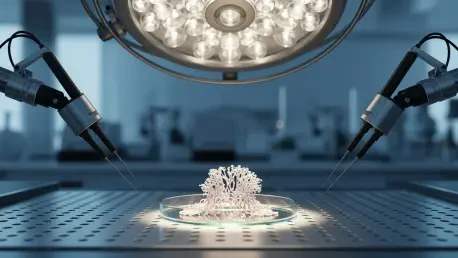

The global medical landscape continues to face a critical shortage of donor organs, leaving millions of patients dependent on invasive treatments like dialysis while they wait for life-saving transplants that may never arrive. For a long time, the promise of growing functional human organs in a laboratory setting seemed like a distant aspiration of science fiction, primarily because researchers struggled to replicate the intricate, multi-layered architecture of natural tissues. While stem cell technology allowed for the creation of basic cellular clusters, these early organoids lacked the refined structural organization necessary to perform complex physiological functions. Without a way to guide cells into their precise anatomical positions, scientists remained stuck with disorganized balls of tissue that failed to mirror the sophisticated reality of a human kidney. Recent breakthroughs at the University of Southern California have finally bridged this gap, introducing a method that uses synthetic organizers to direct growth with precision.
Addressing the Structural Limitations: Why Early Models Failed
Historically, the development of organoids relied heavily on immersing undifferentiated stem cells within a homogenous chemical environment designed to trigger specific biological pathways. Although this approach was successful in producing the basic building blocks of kidney tissue, it fundamentally ignored the dynamic spatial relationships that exist within a developing human embryo. In a natural setting, cells do not receive identical signals all at once; instead, they are influenced by a complex gradient of information that dictates where they should migrate and how they should align. Without these directional cues, laboratory-grown tissues often failed to develop the essential structural axes that define a healthy organ. This lack of organization meant that the resulting models were insufficient for studying the progression of chronic diseases or for conducting reliable pharmacological testing. Consequently, the scientific community recognized that creating a truly functional organ required more than just the right cells.
The absence of a functional collecting duct system remained one of the most significant hurdles in early attempts to bioengineer human kidneys, as these structures are vital for waste transport. Early organoid models were essentially a collection of nephrons without a central plumbing system to connect them, which rendered the tissues physiologically incomplete and structurally erratic. This structural deficiency stemmed from the fact that standard growth protocols could not replicate the specific interaction between the ureteric bud and the surrounding mesenchyme. By failing to establish this critical interface, researchers were unable to trigger the branching morphogenesis that naturally occurs during the first trimester of human development. As a result, the organoids frequently exhibited a disorganized morphology that limited their utility in clinical research and drug discovery. To move toward a solution, it became clear that the environment surrounding the cells needed to be as sophisticated as the cells themselves.
Engineering Precision: The Role of Localized Signaling Hubs
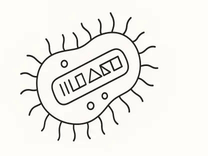
To overcome the chaos of unguided tissue development, researchers introduced synthetic organizers that function as localized signaling hubs within the stem cell population. These engineered cells are designed to secrete specific growth factors in a controlled manner, effectively mimicking the role of natural organizers found in the early stages of human embryogenesis. By strategically placing these hubs within a cluster of stem cells, scientists can create localized gradients of chemical information that encourage the surrounding cells to organize themselves into specific patterns. This method moves away from the traditional top-down approach of flooding a culture with chemicals and instead leverages the innate ability of biological systems to respond to localized environmental cues. The result is a more harmonious developmental process where cells know their position relative to the signaling source, leading to the formation of predictable and anatomically correct structures. This breakthrough represents a fundamental shift in bioengineering.
The success of these synthetic organizers in perfecting kidney architecture established a robust framework for the bioengineering of other complex organs, including the heart and the liver. By proving that localized signaling could dictate the structural integrity of a lab-grown organ, the research community transitioned toward more sophisticated multi-signal models that incorporated vascularization and nerve integration. This advancement moved the medical field significantly closer to the goal of creating patient-specific tissues for drug toxicity screening and disease modeling, which reduced the reliance on animal testing. Future initiatives focused on scaling these organoids into full-sized, transplantable grafts by refining the delivery systems for synthetic signals within larger tissue volumes. It became evident that the future of regenerative medicine lay in the ability to precisely control the spatial environment of stem cells through engineered biological hubs. As these techniques matured, they offered a path toward personalized organ replacements.