
The high cost of exogenous growth factors like BMP-2 remains a major financial barrier to the commercial production of living, engineered bone substitutes. For decades, the reliance on these expensive proteins has restricted high-end tissue engineering to academic research, but the landscape is rapidly shifting toward industrialization. As of 2026, the focus has moved toward a unified production pipeline capable of creating large-scale, mineralized bone constructs that are both economically viable and biologically robust. This advancement represents a significant departure from older, fragmented laboratory techniques that often failed to produce constructs larger than a few millimeters. By integrating high-throughput 3D printing with advanced bioreactor technologies, researchers are developing a blueprint for off-the-shelf bone substitutes. These engineered tissues aim to eliminate the need for autografts, which currently require patients to undergo a separate, painful procedure to harvest bone from their own hip or ribs. The transition to an industrial-ready framework ensures that these complex biological treatments can be standardized and manufactured at a scale that meets the growing demand for orthopedic repairs. This shift is not just about scientific discovery but about creating a sustainable medical supply chain for regenerative therapies that can be deployed across global healthcare systems without the prohibitive price tags of the previous decade.
Hybrid Manufacturing: Achieving Structural Stability
One of the most persistent hurdles in bone tissue engineering is the structural instability of collagen, which is the primary organic component of natural bone. While collagen offers unparalleled biocompatibility and the ability to integrate seamlessly with host tissue, its mechanical weakness makes it notoriously difficult to 3D print into large or geometrically complex shapes. Direct bioprinting techniques frequently result in collapsing structures or require the addition of synthetic cross-linkers that can negatively alter cell behavior or create significant material waste. To address this, a hybrid manufacturing strategy has been implemented that prioritizes both precision and sustainability. Instead of printing the delicate collagen directly, rigid polylactic acid (PLA) male molds are 3D-printed and used to create reusable female molds made of platinum-cured silicone. This indirect approach allows for the casting of a rat-tail type I collagen gel into specific, large-scale architectures that would be impossible to achieve through traditional extrusion printing. By utilizing reusable silicone molds, the process aligns with sustainability principles, reducing the consumption of single-use plastics while ensuring that every scaffold produced has a consistent and tailored geometry.
The resulting collagen scaffolds undergo a specialized process of lyophilization and cross-linking with glutaraldehyde vapor, which imparts a remarkable level of mechanical resilience. These constructs exhibit a unique shape-memory quality, allowing them to be compressed significantly during the cell-seeding process and then spring back to their original dimensions within minutes of being placed in a liquid medium. This physical durability is crucial for the successful populating of the scaffold with human cells, as it ensures the structural integrity of the construct is not compromised during handling. The design of these scaffolds often includes complex internal geometries, such as varying sectional volumes, which allow researchers to study how different physical spaces influence cellular activity within the same unit. This level of structural control is a major step forward from the simple porous blocks of the past, providing a sophisticated framework that can mimic the macro-architecture of a specific patient’s bone defect. By ensuring that the scaffold remains stable and architecturally accurate throughout the entire growth process, the foundation is laid for a bone substitute that can eventually handle the mechanical loads of the human body once it has fully matured in the lab.
Biological Scaling: Achieving Cost-Effective Growth
Scaling up the production of human bone requires an immense number of cells, creating a significant biological bottleneck for clinical applications. Traditional culture methods, such as static T-flasks, are labor-intensive and rarely yield the millions of human bone marrow-derived mesenchymal stromal cells (hMSCs) necessary for large-scale constructs. To solve this, researchers have transitioned to a dynamic expansion system utilizing 250-milliliter spinner flasks. These flasks employ rod impellers and microcarrier beads to create a massive surface area for cell growth within a suspended environment. To prevent the cells from being damaged by fluid forces, computational fluid dynamics (CFD) simulations are used to precisely map the relationship between impeller speed and shear stress. This allows for the identification of an optimal speed, such as 90 revolutions per minute, which maximizes cell yield while maintaining an environment conducive to growth. Through this dynamic approach, it is possible to harvest nearly 20 million cells from a single flask, representing a fourfold increase in efficiency over traditional methods. This high-density expansion ensures that the resulting cells retain their essential biological markers and stemness, remaining ready to transform into bone, cartilage, or fat cells as needed for the final construct.
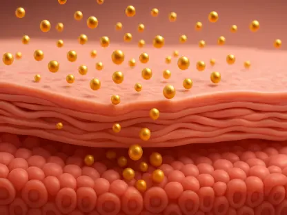
Beyond the sheer quantity of cells, the cost of directing their development into bone has long been a barrier to commercialization. Most engineering protocols require a constant and expensive supply of external growth factors to trigger the necessary biological pathways. A breakthrough in sustainability has been achieved by embedding Chondroitin Sulfate A (CSA), a naturally occurring glycosaminoglycan, directly into the collagen matrix. This creates a biochemical trap that binds and stabilizes the growth factors that the cells naturally secrete themselves, effectively recycling the body’s own signaling mechanisms. This approach mimics the natural process of endochondral ossification, the pathway the body uses to heal major fractures and grow long bones. By utilizing this internal signaling, the need for exogenous proteins is virtually eliminated, and the concentration of other chemical triggers like dexamethasone can be reduced by a factor of 100. This method not only slashes production costs but also ensures more uniform development throughout the interior of large scaffolds, as the signaling molecules are generated by the cells located deep within the tissue rather than being applied to the surface. This self-sustaining biological model is key to making engineered bone a viable option for widespread clinical use.
Perfusion Cultivation: The Pathway to Mineralization
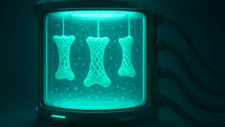
To transform a cell-seeded scaffold into a fully mineralized bone substitute, long-term cultivation in a perfusion bioreactor is required. Over a 60-day period, these constructs are housed within an agarose bedding that protects the delicate collagen from the mechanical forces of fluid flow while simulating the environment of surrounding connective tissue. Inside the bioreactor, growth medium is pumped at a constant rate through channels running parallel to the constructs, ensuring that nutrients and oxygen reach the center of the large scaffolds. This constant flow is a critical improvement over static culture methods, where oxygen levels often drop to dangerous levels in the core of the tissue, leading to cell death and structural collapse. Fiber-optic oxygen sensors are used to monitor the internal environment, confirming that the bioreactor maintains an oxygenated niche that is highly favorable for bone formation. This controlled environment allows the cells to thrive and progress through their developmental stages without the risks associated with stagnant nutrient supplies. The use of real-time monitoring ensures that any deviations in the culture conditions can be addressed immediately, maintaining the stability of the biological process over the several months required for tissue maturation.
The results of this dynamic cultivation process demonstrated a clear superiority over traditional static growth methods. After 60 days, the constructs grown in the perfusion bioreactor maintained their structural integrity and original dimensions, while static samples often suffered from severe shrinkage and internal decay. High-resolution imaging and staining revealed that the perfused scaffolds were mineralized homogeneously throughout their entire volume, reaching densities comparable to natural bone. In contrast, the lack of nutrient flow in static cultures resulted in sparse mineralization that was confined only to the outer edges of the scaffold. Molecular analysis confirmed that the cells followed a precise chronological sequence of biomarkers, starting with cartilage-related genes and ending with the expression of mature bone markers like Runx2 and ALP. This sequence proved that the combination of CSA-enriched scaffolds and perfusion cultivation successfully steered the cells through the complex endochondral ossification pathway. By achieving uniform mineralization in a large-format construct, this research provided the first viable evidence that industrial-scale production of living bone is possible. The ability to control the developmental route of the cells so precisely marks a significant milestone in the field of regenerative medicine.
Future Innovations: Implementing Digital Precision
Looking toward the next phase of this technology, the integration of computational digital twins will play a vital role in optimizing the manufacturing process. These digital models can simulate the bioreactor environment in a virtual space, allowing engineers to predict how cells will react to different flow rates or nutrient concentrations before a single physical experiment is conducted. This predictive capability will further reduce the time and resources needed to tailor bone substitutes for specific patient needs, making the production pipeline even more efficient. Additionally, future iterations of these constructs will incorporate endothelial cells to create internal vascular networks. The presence of these blood vessel precursors will ensure that the bone substitute can quickly integrate with the patient’s circulatory system once implanted, increasing the speed of healing and the overall success rate of the graft. This focus on pre-vascularization addresses one of the final remaining challenges in the transplantation of large tissue-engineered constructs. By combining these advanced computational tools with refined biological techniques, the field is moving toward a standard of care where customized, living bone can be produced on demand from 2026 into 2029 and beyond.
The successful creation of these mineralized constructs established a robust framework for the future of orthopedic medicine. By demonstrating that high-throughput 3D printing and cost-effective biological signaling can produce bone of clinical quality, the study offered a clear path away from the limitations of autologous grafts. The transition from expensive, growth-factor-dependent protocols to sustainable, cell-driven processes represented a major leap in making regenerative therapies accessible to a broader population. Clinicians and researchers can now look toward a reality where complex skeletal defects are treated with precision-engineered tissues that match the patient’s unique anatomy. This industrial-ready pipeline provided the necessary evidence that biological mimicry and engineering precision can work in harmony to solve some of the most difficult challenges in modern surgery. As these technologies continue to evolve, the emphasis will remain on refining the scalability and reliability of the production process to ensure consistent outcomes for every patient. The progress made during this period laid the essential groundwork for a new era of sustainable healthcare, where the laboratory and the clinic are more closely integrated than ever before. Future efforts will focus on clinical trials and regulatory approvals to bring these solutions into standard surgical practice.