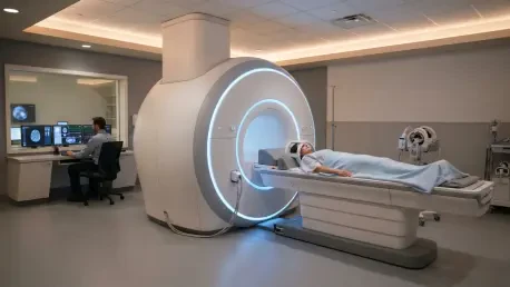

The emergence of contrast-enhanced MRI provides a level of diagnostic granularity that is particularly critical for patients who have a significant family history of disease. For decades, the global medical community relied almost exclusively on mammography as the cornerstone of early cancer detection, but the limitations of this approach became increasingly apparent as diagnostic technology advanced into the mid-2020s. Today, in 2026, the oncological landscape has shifted toward a more nuanced, individualized methodology that prioritizes high-resolution imaging for those most at risk. This evolution is driven by the realization that a one-size-fits-all screening protocol often misses the subtle biological signals of early-stage malignancy. By integrating magnetic resonance imaging into standard clinical workflows, providers have significantly enhanced their ability to identify aggressive tumors before they reach a palpable size, marking a new era of precision medicine and proactive health management for vulnerable populations across the country.
Addressing the Constraints: The Limitations of Standard Mammography
Standard mammography operates through the attenuation of X-rays by various breast tissues, a method that inherently struggles with the differentiation of overlapping structures. In women with high breast density, the fibrous and glandular tissues appear as opaque white regions on the film, which is the same visual signature produced by many cancerous tumors. This visual overlap creates a significant masking effect, where a potentially life-threatening lesion is effectively hidden behind a curtain of healthy, dense tissue. Clinical data suggests that in these specific scenarios, traditional imaging might fail to detect up to half of existing malignancies. This diagnostic blind spot led researchers and clinicians to seek alternative modalities that do not rely on structural density alone but instead focus on the physiological behavior of breast tissue to provide a clearer and more definitive diagnostic picture for patients who require more than just a basic screening tool.
Dense breast tissue is not merely a technical obstacle for radiologists; it is recognized as a significant independent risk factor for the development of breast cancer itself. This reality has necessitated a shift in screening paradigms, moving away from age-based universal standards to risk-based strategies that account for biological variables. While nationalized screening programs have remained robust through recent global healthcare challenges, they often lack the specificity required for younger women who exhibit higher tissue density or those with known genetic predispositions. For these individuals, the gap between standard care and effective surveillance was once wide, often leading to delayed diagnoses that necessitated more aggressive treatment paths. The integration of advanced magnetic resonance imaging has begun to bridge this divide, offering a reliable safety net for populations that mammography fails to protect by providing a clear view through even the densest glandular tissue.
Biological Beacons: Visualizing Cancer through Contrast Agents
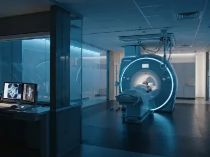
The technical superiority of advanced breast MRI is rooted in its ability to generate high-resolution, three-dimensional visualizations of the breast anatomy without the use of ionizing radiation. Instead, the process utilizes powerful magnetic fields and radiofrequency pulses to excite hydrogen atoms within the body’s tissues, capturing the resulting signals to construct detailed cross-sectional images. The inclusion of a contrast agent, typically a gadolinium-based compound, elevates this process from a structural scan to a functional diagnostic tool. When this agent is administered intravenously, it permeates the entire circulatory system, allowing radiologists to observe the real-time dynamics of blood flow within the breast. This approach provides a level of clarity that ultrasound or X-ray cannot match, as it highlights functional abnormalities that occur at the cellular and microvascular levels long before structural changes become visible to the human eye.
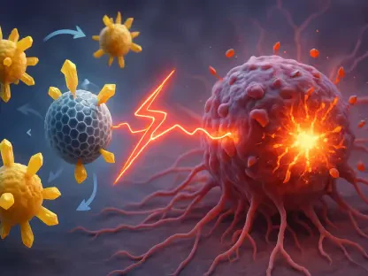
The biological foundation for using contrast-enhanced imaging is a process known as angiogenesis, which is the formation of new blood vessels that malignant tumors require to sustain their rapid growth. These tumor-induced vessels are structurally defective, characterized by a higher degree of permeability or leakiness compared to the organized vasculature found in healthy mammary tissue. As the gadolinium contrast agent moves through the bloodstream, it leaks out of these irregular vessels and accumulates in the surrounding tumor tissue, creating a bright signal on the MRI scan. This phenomenon allows clinicians to detect even microscopic clusters of cancerous cells by identifying their metabolic and vascular signatures. By focusing on the physiological demand of a growing tumor rather than just its shape or density, contrast-enhanced MRI identifies suspicious areas with a sensitivity that was previously unattainable in general clinical practice.
Strategic Deployment: Targeted Surveillance and Surgical Planning
Rather than replacing mammography for the general population, contrast-enhanced MRI is strategically deployed for individuals who fall into specific high-risk categories. Clinical guidelines established by 2026 emphasize the use of this technology for women carrying BRCA1 or BRCA2 mutations, whose lifetime risk of developing disease is exponentially higher than the average. Additionally, patients with a documented history of chest radiation therapy or those with rare syndromes like Li-Fraumeni require this level of surveillance. For these groups, the annual screening regimen now typically includes a combination of mammography and MRI, ensuring that any new developments are caught at the earliest possible stage. This targeted approach ensures that healthcare resources are directed where they provide the most significant impact, offering a proactive defense for patients who face a statistically higher probability of facing a difficult diagnosis.
In the context of a confirmed diagnosis, MRI has become indispensable for the preoperative planning and staging of the disease. It allows the surgical team to map the precise dimensions and location of a tumor within the breast, which is vital for achieving successful outcomes in breast-conserving surgeries. Often, the high sensitivity of the MRI reveals satellite lesions or multifocal disease that remained hidden on other imaging modalities, preventing the need for secondary surgeries and ensuring that the initial treatment plan is comprehensive. This precise mapping also aids in the evaluation of the contralateral breast, ensuring that no concurrent malignancies go unnoticed. Leading medical centers have integrated these MRI findings into multidisciplinary tumor board discussions, where oncologists, surgeons, and radiologists collaborate to design a personalized treatment path that minimizes recurrence and maximizes safety.
Clinical Realities: Navigating Challenges and Future Breakthroughs
Despite its profound diagnostic advantages, the extreme sensitivity of MRI introduces a unique set of clinical challenges, most notably the high rate of false positives. The technology is so effective at identifying minute variations in tissue that it often flags benign conditions, such as fibroadenomas or hormonal changes, as potentially suspicious. These findings frequently necessitate follow-up biopsies, which can cause significant emotional and psychological stress for patients who are already navigating a high-risk landscape. Furthermore, the procedural requirements of an MRI can be taxing, as patients must remain completely motionless for up to 45 minutes in a prone position. For those with claustrophobia or certain musculoskeletal issues, the experience can be difficult to tolerate. Additionally, the use of gadolinium requires careful screening of renal function to prevent rare systemic complications.
The diagnostic field is currently moving toward more efficient and patient-friendly solutions by implementing abbreviated MRI protocols. These fast MRI techniques focus on capturing the most critical contrast-enhanced sequences, reducing the total time a patient spends in the machine from nearly an hour to under fifteen minutes. This reduction not only improves the patient experience but also decreases the cost per scan, making the technology accessible to a wider demographic. Simultaneously, the integration of Artificial Intelligence is transforming the way radiologists interpret these complex data sets. Advanced algorithms are now capable of filtering through thousands of images to highlight regions of interest, reducing human error and helping to distinguish between benign enhancements and actual malignancies. These technological advancements are working in tandem to refine the balance between high sensitivity and clinical specificity.
Past Progress: Developing a Personalized Diagnostic Roadmap
The transition toward a more nuanced application of contrast-enhanced MRI marked a significant milestone in the history of oncological diagnostics. Healthcare systems successfully pivoted from rigid, population-wide screening models to a more agile, risk-stratified approach that saved countless lives through earlier detection. This shift necessitated a major investment in both technological infrastructure and specialized training for medical professionals, ensuring that the granularity provided by MRI was translated into actionable clinical results. Patients and providers alike benefited from the increased clarity, which reduced the ambiguity that often surrounded the screening of high-risk individuals. By addressing the limitations of previous imaging standards, the medical community established a new baseline for what constitutes effective cancer surveillance, paving the way for a diagnostic environment where specific needs were met with precision.
The long-term success of these imaging initiatives depended on the integration of genetic counseling and personalized risk assessment into the routine care of every patient. Medical teams learned to utilize the high-resolution data from MRI not just for detection, but as a roadmap for holistic health management that included preventive measures and lifestyle interventions. This comprehensive strategy proved to be the most effective way to manage the complexities of hereditary disease and high tissue density. As imaging technologies continued to become more streamlined and affordable, the barriers that once prevented widespread adoption began to dissolve, allowing for a more equitable distribution of advanced care. Ultimately, the evolution of breast MRI demonstrated that the most effective way to combat complex health challenges was through the continuous refinement of diagnostic tools and the commitment to treating every patient uniquely.