
While the human eye has long been revered as the window to the soul, contemporary medical breakthroughs are establishing that its intricate structures provide an even more profound map of a patient’s cognitive longevity. Traditionally, a diagnosis of Alzheimer’s disease arrived far too late, often only after irreversible brain damage had already settled into the neural tissue and compromised the patient’s daily life. This delayed reality has historically left families with few options beyond palliative care, but the emergence of artificial intelligence in ophthalmology is fundamentally altering this trajectory. By repurposing the standard retinal scan, medical science now offers a non-invasive and highly accessible path to identifying cognitive decline long before the first signs of memory loss appear.
This transformation is turning routine eye examinations into high-performance diagnostic tools that do more than just measure vision. The integration of massive datasets with advanced image processing allows clinicians to observe the subtle neurovascular changes that signal systemic health issues. Beyond mere vision correction, these tools represent a shift toward a comprehensive monitoring system for global neurovascular health. This development redefines the role of the optometrist as a vital participant in the broader medical landscape, providing a frontline defense against the silent onset of neurodegeneration.
The Convergence of AI and Ophthalmology
Growth Trends and Data-Driven Diagnostics
The rapid evolution of this technology relies heavily on the shift toward large-scale biobanking, which provides the necessary fuel for training sophisticated neural networks. Data repositories, most notably the UK Biobank, have allowed researchers to analyze over 40,000 detailed retinal records to identify patterns that previously escaped human detection. This transition from subjective patient self-reporting to objective digital analysis represents a significant leap in clinical reliability. While patients might struggle to accurately recall their lifestyle habits or blood pressure history, the retina maintains a physical record of the cumulative biological stress experienced by the body.
Current AI models demonstrate an uncanny ability to predict specific biological traits and lifestyle behaviors with high statistical accuracy through simple imagery. Algorithms can now determine a patient’s sex, cardiovascular pressure, and even history of smoking or chronic insomnia without invasive blood tests or lengthy questionnaires. This wealth of information is extracted by identifying how these factors leave a permanent signature on retinal morphology. As these models become more refined, the reliance on external data points decreases, placing the retinal scan at the center of the modern diagnostic toolkit.
Real-World Applications and Breakthrough Research
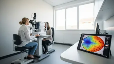
Significant strides in this field have been championed by Dr. Ruogu Fang at the University of Florida, whose research has highlighted the untapped potential of standard ocular imaging. Findings published in the Journal of Alzheimer’s Disease emphasize that the hardware required for these screenings is already present in most modern clinics. By utilizing low-cost retinal photography, healthcare providers are essentially turning a basic vision tool into a sophisticated biological sensor for neurodegeneration. This democratization of screening ensures that early detection is not limited to those with access to elite neurological centers.
The diagnostic focus centers on specific biomarkers, such as the health of retinal arteries and the integrity of the optic nerve. AI scans these structures for microvascular thinning or unusual nerve patterns that correlate with the early stages of Alzheimer’s risk. Because these changes often occur years before clinical symptoms manifest, the window for intervention is significantly widened. Moreover, this research provides the clinical evidence necessary to move these tools out of the laboratory and into the primary care environment where they can do the most good.
Expert Perspectives on the Retinal “Biological Sensor”
The medical community increasingly views the eye as an integrated sensor for the body’s entire neurovascular system, rather than an isolated organ. This conceptual shift has led experts to prioritize early-stage vascular biomarkers over traditional late-stage indicators like amyloid plaques in the brain. Since the retina is embryonically derived from the same tissue as the brain, it serves as a proxy for what is happening within the central nervous system. Experts argue that monitoring these vessels of the mind provides a clearer, more immediate picture of cognitive health than traditional cognitive testing.
Furthermore, the layer of objectivity provided by AI is considered essential for modern diagnostics. Human clinicians, regardless of their expertise, are limited by the resolution of the human eye and the inherent biases of visual interpretation. Artificial intelligence, however, uncovers subtle variations in retinal texture and vessel curvature that are statistically significant but visually imperceptible. This ability to quantify risk with mathematical precision allows for a more standardized approach to neurovascular care, reducing the likelihood of misdiagnosis or overlooked early-stage symptoms.
Future Outlook: Implications for Global Healthcare
The scalability of retinal screening offers a stark contrast to the high costs and logistical hurdles associated with traditional neurological imaging. While MRI and PET scans are often prohibitive due to expense and the need for specialized facilities, retinal photography is fast, affordable, and readily available in suburban and rural areas. This ease of use suggests a future where routine cognitive screenings are as common as checking one’s blood pressure or vision prescription. Such a shift allows for mass screening programs across diverse populations, ensuring that neurodegenerative risks are caught in the earliest possible phases.
Proactive intervention becomes the new standard of care under this model, allowing for personalized lifestyle modifications and specialized medications to be deployed early. However, the path forward is not without challenges, as the integration of these tools into standard clinical workflows requires significant coordination across specialties. Ethical considerations regarding early-stage risk notification also remain a topic of debate, as patients must be prepared for the psychological impact of knowing their long-term cognitive risks. Despite these hurdles, the fusion of AI and ophthalmology is poised to redefine routine vision care as a primary defense against the global Alzheimer’s crisis.
Conclusion: A Proactive Defense for Cognitive Longevity
The transformation of retinal photography into a vital neurological diagnostic tool marked a turning point in the fight against cognitive decline. By shifting the medical focus from late-stage treatment to early-stage prevention, the industry successfully harnessed the power of AI to identify risks that were once invisible. This proactive approach provided a new level of agency for patients, allowing them to take control of their cognitive health before irreversible damage took hold. It established a standard where cognitive longevity became a manageable metric rather than an unpredictable outcome.
Looking ahead, the clinical adoption of these screenings reshaped the landscape of neurodegenerative disease management by prioritizing accessibility. The next logical step involves the integration of these tools into wearable devices and home-monitoring systems, which could allow for continuous neurovascular tracking. By ensuring the defense of the mind is as constant as the monitoring of a heartbeat, medical science offers a future where Alzheimer’s is intercepted long before it can alter a life. This evolution highlights the necessity of adopting AI-driven screening as the gold standard for global healthcare.