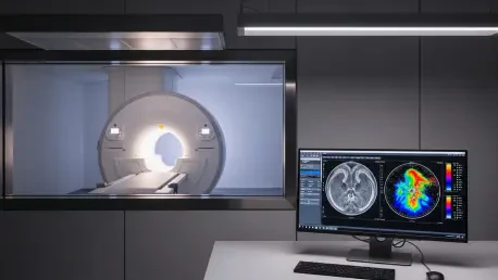

The transition of magnetic resonance imaging from a qualitative art form into a rigorous quantitative science represents one of the most significant shifts in medical diagnostics within the current decade. For many years, clinicians relied on visual interpretations of contrast and shadow, but the arrival of the Titanion MR at the International Society for Magnetic Resonance in Medicine conference has introduced a platform specifically engineered to extract biological data directly from human tissue. This 3.0T system is not simply an evolution of hardware; it serves as a sophisticated bridge that allows high-level academic research into cellular environments to enter the routine clinical environment. By integrating extreme gradient performance with advanced artificial intelligence, this technology provides the necessary precision to monitor the microscopic behaviors of water molecules and metabolites. Such capabilities are essential for identifying the subtle, early-stage biological shifts associated with aggressive cancers and complex neurodegenerative conditions that were previously invisible.
Engineering Precision for Deep Tissue Insights
High-Gradient Performance: The Key to Microstructural Data
The technical foundation of the new system is built upon an unprecedented gradient strength of 150 mT/m coupled with a slew rate of 250 T/m/s, which establishes a new benchmark for 3.0T MRI performance. These specifications are not merely academic milestones; they provide the raw physical power required to perform advanced diffusion-weighted imaging at a scale that was once the exclusive domain of specialized research magnets. High gradient strength allows the system to sensitize the signal to the movement of water molecules over incredibly short distances, revealing the intricate architectural details of cellular membranes and interstitial spaces. This level of detail is paramount when clinicians need to differentiate between healthy tissue and the dense, disorganized cellular growth characteristic of early-stage tumors. By capturing these microscopic nuances, the machine provides a much clearer window into the physiological state of the patient, allowing for a more nuanced understanding of disease progression.
In addition to its raw power, the system is designed to sustain this high level of performance throughout the duration of complex, multi-sequence examinations. The engineering focus on high Grms performance ensures that the gradients can operate at peak capacity without the thermal drift or mechanical fatigue that often limits the effectiveness of high-performance imaging in a high-volume clinical setting. This stability is particularly important for quantitative mapping, where any fluctuation in gradient output can lead to inaccuracies in the calculated biomarker values. By maintaining a consistent magnetic environment, the platform allows for the generation of standardized data sets that can be compared across different scans and different patients. This reliability transforms the MRI from a diagnostic tool into a precision measurement instrument, enabling the medical community to develop more robust longitudinal studies and refine the accuracy of predictive models for various chronic health conditions.
Signal Fidelity: Eliminating Distortions for Precise Measurements
Achieving high-quality quantitative data requires more than just powerful magnets; it demands a radical reduction in the physical artifacts that typically accompany high-speed magnetic switching. The Titanion MR addresses this through a sophisticated low eddy current design, which minimizes the secondary magnetic fields that can blur images and corrupt the integrity of sensitive measurements. Eddy currents are a common byproduct of rapid gradient transitions, and in traditional systems, they often necessitate significant software correction that can unintentionally smooth over critical clinical details. By preventing these distortions at the hardware level, the system ensures that the captured signal remains as close to the original biological source as possible. This high level of signal fidelity is the cornerstone of reliable biomarker development, as it provides a clean and untainted baseline for the complex mathematical algorithms used to calculate tissue density, perfusion, and metabolic activity.
Building on this foundation of hardware stability, the system incorporates advanced shielding and coil technologies that further enhance the signal-to-noise ratio. This improvement is vital when attempting to image deep-seated structures or when using ultra-fast acquisition protocols that naturally tend to lose signal strength. The result is a diagnostic environment where clinicians can trust the numerical output of the machine, whether they are measuring iron deposition in the brain or assessing the vascularity of a suspicious lesion in the abdomen. Furthermore, the reduction in artifacts allows for more precise registration between different imaging modalities and time points, which is essential for tracking the effectiveness of systemic therapies. By prioritizing the purity of the data stream, the platform enables a shift toward evidence-based radiology where every pixel carries a specific, quantifiable meaning that contributes to a deeper understanding of the patient’s internal biological landscape.
Transforming Diagnostic Capabilities Across Specialties
Neurological Advancement: Beyond Anatomical Shrinkage
The move toward quantitative biomarkers is perhaps most impactful in the field of neurology, where traditional imaging often fails to detect the earliest stages of cognitive decline. Historically, clinicians had to wait for visible anatomical shrinkage or significant functional loss to confirm a diagnosis of a neurodegenerative disorder, at which point the damage was often irreversible. The Titanion MR changes this dynamic by allowing for the visualization of tissue microstructure, providing a way to see the biological “wear and tear” at a microscopic level long before it manifests as structural atrophy. By quantifying the diffusion properties of white matter tracts and the metabolic health of grey matter, researchers can identify subtle signs of pathology that indicate a high risk of future decline. This early insight is critical for the development of neuroprotective strategies and allows for the enrollment of patients in clinical trials at a stage when therapeutic interventions are most likely to be effective.
Furthermore, the ability to measure these microscopic changes with high precision supports the discovery of new imaging biomarkers that can track the real-time progression of brain diseases. Instead of relying on subjective assessments of patient symptoms, neurologists can use objective data to monitor how a disease is evolving and how the brain is responding to various treatments. This approach is particularly valuable for complex conditions like multiple sclerosis or traumatic brain injury, where the relationship between clinical presentation and physical damage is not always straightforward. The system’s high-resolution capabilities enable the mapping of small-scale inflammatory processes and changes in blood-brain barrier integrity, providing a comprehensive picture of neurological health. This transition toward data-driven neurology not only improves the accuracy of individual diagnoses but also contributes to a larger body of knowledge that will ultimately lead to more effective and personalized treatment protocols.
Oncology and Whole-Body Scanning: A Comprehensive Framework
In the realm of oncology, the integration of high-resolution microstructural data with a massive 55 cm field of view offers a transformative approach to cancer staging and monitoring. Traditional MRI scans are often limited in scope, requiring multiple separate examinations to assess the spread of a systemic disease across different organ systems. The broad coverage of the Titanion MR allows for a comprehensive whole-body assessment in a single session, ensuring that no potential metastatic site is overlooked. This holistic view is essential for determining the most appropriate course of treatment, as it provides a clear picture of the total tumor burden and its distribution throughout the body. By combining this wide-angle perspective with the ability to perform deep-tissue quantitative analysis, the system allows oncologists to characterize the biological aggressiveness of different lesions simultaneously, leading to more informed and timely clinical decisions.
This comprehensive framework also facilitates more accurate monitoring of a patient’s response to modern systemic therapies, such as immunotherapy or targeted molecular agents. Because these treatments can cause complex biological reactions that do not always result in immediate tumor shrinkage, traditional size-based measurements can be misleading. The quantitative biomarkers provided by this platform allow for the assessment of changes in cellularity, vascularity, and metabolic activity, which often precede structural changes. This enables a “functional” assessment of treatment efficacy, allowing clinicians to pivot away from ineffective therapies much sooner and transition patients toward more successful options. This shift toward longitudinal, data-rich monitoring is a vital component of personalized oncology, where the goal is to tailor every aspect of care to the unique biological profile of the individual patient and their specific disease, thereby improving long-term outcomes and quality of life.
Optimizing Clinical Workflows and Infrastructure
Artificial Intelligence: Enhancing Speed Without Sacrificing Quality
One of the most persistent challenges in magnetic resonance imaging has been the inherent trade-off between the time required for a scan and the quality of the resulting images. The introduction of the SmartSpeed Precise AI suite directly addresses this conflict by using sophisticated deep-learning algorithms to reconstruct high-fidelity images from significantly less raw data. This allows the system to produce scans that are up to 80% sharper than conventional images while simultaneously reducing the total time a patient spends inside the bore by a factor of three. This reduction in scan time is a critical improvement for patient comfort, particularly for those who suffer from claustrophobia or have difficulty remaining still for long periods. By making the imaging process faster and less stressful, the technology ensures a higher success rate for examinations and reduces the need for repeat scans due to patient movement or discomfort, which is a major win for hospital efficiency.
The increase in throughput made possible by these AI-driven workflows also has profound implications for the operational economics of modern healthcare facilities. Hospitals can now serve a much larger number of patients each day without having to invest in additional hardware or expand their physical footprint. Moreover, the enhanced image clarity provided by the AI algorithms gives radiologists greater confidence in their interpretations, leading to faster and more accurate reporting. This streamlined workflow reduces the diagnostic bottleneck that often occurs in busy medical centers, allowing patients to move through the care continuum more quickly. By automating the most labor-intensive aspects of image reconstruction and optimization, the system frees up medical professionals to focus more on patient interaction and complex clinical analysis. This synergy between human expertise and machine intelligence represents the future of medical imaging, where technology acts as a force multiplier for clinical excellence.
Infrastructure Compatibility: Decentralizing High-Performance Care
Historically, the adoption of ultra-high-performance MRI technology was restricted to a handful of elite academic institutions due to the massive power and cooling requirements of such systems. Many high-gradient machines required dedicated electrical substations and extensive architectural modifications to accommodate their heavy infrastructure needs. The Titanion MR breaks this trend by operating with a relatively modest power requirement of only 115 kVA, which is comparable to many standard 3.0T systems already in use. This engineering feat allows hospitals and regional clinics to upgrade their diagnostic capabilities without the prohibitive costs of a major facility overhaul. By lowering the barriers to entry, the technology facilitates the democratization of advanced imaging, ensuring that patients in a wider variety of geographic locations can access the benefits of high-end quantitative biomarkers and microstructural diagnostics.
This focus on operational scalability is essential for moving advanced research-grade technology into routine clinical practice across the global healthcare landscape. When high-performance tools are easier to install and maintain, they can be deployed in community hospitals and specialized outpatient centers where the majority of patient care actually takes place. This decentralization of technology helps to reduce disparities in care, as patients no longer need to travel to major metropolitan hubs to receive the most advanced diagnostic assessments. Furthermore, the standardized nature of the quantitative data produced by these systems ensures that clinical insights remain consistent regardless of where the scan was performed. This interoperability is vital for large-scale health initiatives and collaborative research, as it allows for the seamless integration of data from diverse clinical settings into a unified framework for improving global health outcomes and developing new medical standards.
Strategic Pathways: The Future of Quantitative Health
The introduction of the Titanion MR marked a decisive moment in the evolution of medical diagnostics, as the focus transitioned from visual observation to data-driven precision. By successfully merging extreme hardware performance with intelligent software, the system provided a clear roadmap for the future of personalized medicine. Clinical teams recognized that the ability to quantify biological processes across the entire body was not just an incremental improvement but a fundamental shift in how diseases were managed and understood. The integration of high-fidelity signal processing and low infrastructure requirements demonstrated that elite diagnostic power could be made accessible to a broader range of facilities, effectively moving the cutting edge of research into the hands of daily practitioners. This move toward standardized, objective measurement laid the groundwork for a more predictive and proactive healthcare model that prioritized early intervention and tailored treatment plans.
Moving forward, the medical community should prioritize the integration of these quantitative imaging biomarkers into standardized clinical pathways to fully realize the potential of this technology. Healthcare organizations must invest in training for radiologists and technicians to ensure they can leverage the full suite of AI-enhanced tools and microstructural analysis techniques. Furthermore, longitudinal data collection should be emphasized to refine the predictive accuracy of the biomarkers generated by these high-performance systems. By focusing on the continuous improvement of data quality and the expansion of clinical applications, the industry can ensure that every MRI scan provides the maximum possible insight into patient health. This proactive stance will accelerate the transition toward a healthcare system that is defined by its ability to see and treat the earliest signs of disease, ultimately leading to improved survival rates and a better quality of life for patients across all medical specialties.


