
The insidious nature of glaucoma often means that permanent vision loss occurs long before a patient even realizes that their ocular health is in significant and immediate danger. For decades, the medical community has struggled with the reality that although this condition is the leading cause of irreversible blindness, many individuals remain undiagnosed until extensive damage is done. Traditional screening methods, while accurate, are frequently inaccessible to the broader population due to the massive physical footprint and high maintenance costs of standard ophthalmic equipment. This gap in the healthcare system creates a pressing need for a diagnostic tool that is not only highly precise but also sufficiently portable to reach patients in remote or underserved environments. PeriVision, a specialized medical technology firm emerging from the University of Bern, is actively bridging this divide by integrating high-precision Swiss engineering with advanced artificial intelligence. By rethinking the mechanics of visual field testing, the company is transforming a traditionally grueling clinical procedure into a streamlined, patient-centric experience that promises to democratize specialized eye care on a global scale.
Academic Foundations: Bridging Research and Commercial Reality
The foundation of PeriVision was established through a rigorous synergy between sophisticated commercial strategies and deep academic research conducted at the University of Bern. This specific multidisciplinary origin allowed the company to leverage years of laboratory-tested vision data to create tools that are not only theoretically sound but also practically applicable in busy clinical environments. By assembling a leadership team that combines expertise in medical imaging, artificial intelligence, and international business, the firm successfully navigated the complex path from a scientific prototype to a viable medical device. This unique blend of skills ensured that every technological advancement was grounded in clinical evidence while remaining scalable for a global market. The commitment to maintaining a high standard of Swiss engineering meant that the hardware components were designed for durability and precision, addressing the specific needs of ophthalmologists who require reliable data to make critical decisions regarding surgery or medication.
This academic pedigree provided the necessary credibility to attract investors and clinical partners who were wary of unproven technology in the sensitive field of ophthalmology. The developers focused on creating a platform that could handle complex vision data without requiring a team of technicians to operate it constantly. This transition from a university-bound research project to a standalone commercial entity was marked by a series of rigorous trials that confirmed the platform’s diagnostic accuracy compared to industry gold standards. By maintaining strong ties with the scientific community, the company ensured that its AI models remained at the cutting edge of what is possible in vision science. The objective was clear: to create a solution that offers the high-level performance of a specialized hospital department in a format that could be utilized by general practitioners and local clinics. This approach effectively minimized the distance between advanced laboratory breakthroughs and the patients who need them most in their daily lives.
The Diagnostic Crisis: Addressing Accessibility and Patient Fatigue
The global burden of visual impairment is staggering, with glaucoma alone affecting approximately 80 million individuals, a number that continues to grow as the population ages. While many cases are manageable if identified during the early stages, current diagnostic systems often fail to facilitate widespread screening. Traditional machines are typically large, stationary, and require a significant financial investment, making them difficult to deploy outside of specialized urban hospitals. This lack of accessibility means that patients in rural or low-income areas often miss the critical window for intervention. Furthermore, the high cost of maintenance for these legacy systems places a heavy financial burden on clinics, which often results in higher costs for patients. Without a more flexible and cost-effective alternative, the medical community would continue to struggle with a backlog of patients who require regular monitoring to prevent the total loss of their sight.
Beyond the logistical hurdles, the standard testing process itself is often physically and mentally taxing for patients who must maintain absolute focus for long periods. During a procedure known as perimetry, patients are required to stare at a fixed point while responding to flashes of light, a task that quickly leads to fatigue and inconsistent results. This exhaustion is not just a comfort issue; it directly impacts the accuracy of the diagnostic data, as tired patients are more likely to miss stimuli or provide false positives. Consequently, clinical staff must spend excessive time supervising each test to ensure the validity of the results, creating significant bottlenecks in patient flow. These challenges highlight the need for a diagnostic method that reduces the cognitive load on the patient while simultaneously freeing up medical personnel to focus on other aspects of care. The current reliance on antiquated, high-pressure testing environments is no longer sustainable for a global healthcare system.
Algorithmic Precision: The Impact of Predictive Machine Learning
To overcome these significant hurdles, the engineering team developed the Sequential Optimization Reconstruction Strategy, commonly referred to as the SORS algorithm. This proprietary AI-driven approach fundamentally changes how visual field testing is conducted by moving away from the traditional method of testing every single point on a predefined grid. Instead, the system utilizes advanced machine learning models trained on thousands of clinical visual fields to identify the most critical points for each individual. By testing a small, strategically selected subset of locations, the algorithm can predict the sensitivity of the remaining areas with an incredibly high degree of mathematical accuracy. This shift from exhaustive testing to predictive modeling represents a major leap forward in diagnostic efficiency, ensuring that no time is wasted on redundant data collection while still maintaining the clinical integrity required for a diagnosis.
The efficiency gains provided by this predictive modeling are nothing short of revolutionary, reducing the total time required for a comprehensive test by approximately 70 percent. While industry-standard tests can often take over ten minutes per eye, the SORS algorithm allows for the completion of the same task in just two to three minutes. Clinical data suggests that this dramatic increase in speed does not compromise the reliability of the results; in fact, the system often produces cleaner data by finishing the test before the patient begins to experience significant exhaustion. By minimizing the duration of the procedure, clinics can process a much larger volume of screenings without expanding their physical footprint or hiring additional staff. This capability is essential for managing the growing number of patients who require frequent monitoring to track the progression of their condition. The algorithm effectively turns a time-consuming chore into a rapid and reliable clinical assessment.
Portable Solutions: Virtual Reality in Clinical Environments
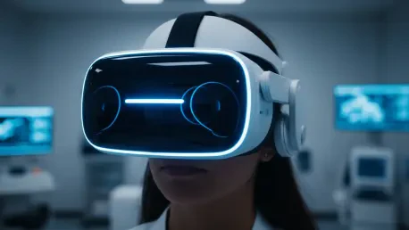
The delivery of this advanced technology is made possible through a lightweight, 0.6-kilogram virtual reality headset that serves as a portable visual field detector. This device effectively eliminates the need for dedicated clinical darkrooms and bulky, desk-mounted equipment that takes up valuable office space. Because the headset creates its own controlled lighting environment, it can be used in almost any setting, from a standard exam room to a mobile clinic or even a patient’s home. The inclusion of a built-in virtual assistant provides automated guidance in multiple languages, which allows patients to complete their examinations with very little direct supervision from medical personnel. This automation is a key feature for clinics looking to optimize their workflow, as it allows technicians to manage multiple screenings simultaneously without sacrificing the quality of the patient experience or the accuracy of the data.
To further ensure the integrity of the collected information, the headset is equipped with active eye-tracking sensors that monitor the patient’s focus in real-time. If a patient’s gaze wanders from the central fixation point, the system can pause the test or adjust the data to account for the movement, preventing the errors that frequently plague traditional perimetry. Furthermore, the system utilizes adaptive testing intervals that adjust the speed of light stimuli based on the patient’s individual reaction time. This personalization makes the test feel less mechanical and more responsive, which significantly improves the overall patient experience. These features collectively increase clinic throughput by up to 30 percent while reducing the total labor time required from staff by over 60 percent. The result is a more human-centric approach to medical technology that prioritizes both the operational needs of the provider and the physical comfort of the individual undergoing the test.
Global Reach: Regulatory Success and Market Expansion
The transition of this platform from a specialized tool to a global standard was accelerated by securing necessary regulatory certifications in the United States, the United Kingdom, and the European Union. These approvals cleared the path for widespread international use, allowing healthcare providers across different continents to integrate the VR-based system into their standard diagnostic workflows. A successful recent seed-funding round of 2.4 million Swiss francs provided the necessary capital to expand the platform’s capabilities beyond its initial focus. Moving forward, the strategic plan involved adapting the virtual reality technology to assist in the diagnosis of other retinal diseases, such as age-related macular degeneration. This expansion into broader ophthalmic applications ensured that the hardware remained a versatile asset for clinics, capable of performing multiple diagnostic functions through simple software updates and continued AI refinement.
The successful implementation of these diagnostic protocols showed that the integration of virtual reality and machine learning was essential for modernizing ophthalmic care. Healthcare administrators who adopted these systems observed that the reduction in manual labor allowed staff to focus on complex patient cases rather than routine testing supervision. The expansion into the Chinese market demonstrated the scalability of the technology across diverse clinical settings and varied economic landscapes. By finalizing the seed-funding round, the organization secured the necessary resources to explore therapeutic applications for other retinal conditions, effectively moving beyond mere detection into holistic management. These advancements established a clear precedent for decentralized medical testing, suggesting that the future of specialized care must involve tools that are as portable as they are intelligent. Stakeholders ensured that the continuous improvement of the predictive algorithms remained a priority to maintain diagnostic accuracy.