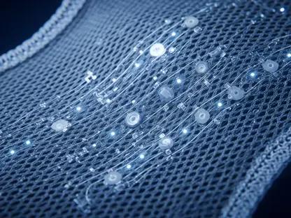

Within the immensely complex and crowded biological environment of the human anatomy, approximately thirty-seven trillion individual cells are constantly engaged in a silent, microscopic dialogue that dictates the fundamental parameters of health and disease. This intricate communication network relies on exosomes, which are tiny, membrane-bound vesicles that act as biological cargo ships. These extracellular vesicles travel through the bloodstream and other bodily fluids, transporting a concentrated payload of proteins, lipids, and genetic material from their parent cells to distant targets. By studying these messengers, scientists can essentially eavesdrop on the internal state of the body, potentially uncovering early warning signs of illness long before physical symptoms appear.
The pursuit of decoding these cellular transmissions has become a central objective in the field of non-invasive medicine. The promise is profound: the ability to diagnose cancer, neurodegenerative disorders, or autoimmune diseases through a simple blood draw. Because exosomes carry the specific molecular “fingerprint” of the cells that produced them, they offer a direct window into the pathological processes occurring in deep tissues. However, the sheer smallness of these particles has historically kept them just out of reach for traditional diagnostic equipment, leaving their vital messages largely unread. Modern researchers now recognize that mastering exosome analysis is the key to unlocking the next generation of precision healthcare and liquid biopsies.
Beyond the Microscope: Unveiling the Secret Language of 37 Trillion Cells
The role of exosomes in systemic health is far more significant than previously understood, as these vesicles facilitate the transfer of functional molecules that can alter the behavior of recipient cells. In the context of oncology, for instance, tumors use exosomes to prepare distant sites for metastasis, essentially “priming the soil” before the cancer cells arrive. Similarly, in the brain, these vesicles carry proteins associated with Alzheimer’s and Parkinson’s, serving as both a mechanism for disease spread and a potential marker for its progression. The discovery that these particles are not merely cellular waste but active participants in biological regulation has fundamentally shifted the landscape of medical research.
Despite their importance, the microscopic nature of exosomes—often measuring less than 100 nanometers in diameter—makes them difficult to isolate and characterize. To put their size in perspective, they are approximately one-thousandth the width of a human hair. This physical limitation means that conventional optical microscopes simply cannot resolve the details of an individual exosome. Consequently, much of the early work in this field focused on the collective behavior of these particles, rather than the unique data points held within a single vesicle. As the scientific community moved toward more personalized treatment models, the need for a tool capable of seeing individual exosomes became an urgent priority.
The Diagnostic Bottleneck: Why Traditional Exosome Research Hit a Wall
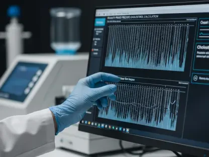
The primary obstacle in current exosome research is the reliance on “bulk” analysis, a method that measures the average characteristics of millions of vesicles simultaneously. While this provides a general overview, it frequently overlooks the rare, disease-carrying exosomes that might be hidden within a sea of healthy ones. This lack of sensitivity is particularly problematic for early-stage cancer detection, where the “signal” from a small tumor might be completely drowned out by the noise of normal cellular activity. Furthermore, traditional techniques like nanoparticle tracking analysis or electron microscopy provide structural data but often fail to deliver the chemical and molecular context required for a definitive diagnosis.
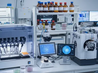
In addition to the resolution issue, the existing diagnostic workflow is hindered by a labor-intensive and time-consuming process. Isolating exosomes from a clinical sample usually requires multiple rounds of high-speed centrifugation and complex purification steps that can take days to complete. Many current analytical methods also necessitate large sample volumes and sophisticated DNA or protein amplification, which adds layers of cost and potential error. These inefficiencies have created a significant gap between the promising results seen in research laboratories and the practical needs of a fast-paced clinical environment. Without a more streamlined and precise method for single-vesicle profiling, the potential of exosome-based diagnostics remained largely untapped.
Inside INSPECT: A Tri-Modal Leap in Single-Exosome Analysis
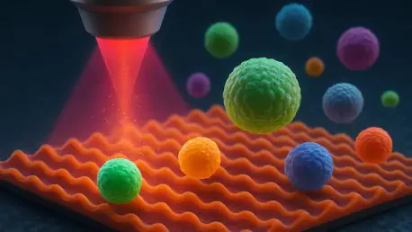
The University of Houston has pioneered a technological solution to these challenges with the development of the Integrated Nanophotonic Imaging and Spectroscopy Technology, or INSPECT. This platform represents a significant departure from conventional methods by utilizing nanoplasmonic enhancement to study exosomes on an individual basis. By focusing light into tiny volumes at the nanoscale, the INSPECT system can amplify signals that would otherwise be too weak to detect. This allows researchers to bypass the need for bulk averaging and instead focus on the specific molecular makeup of each particle, providing a level of granularity that was previously impossible to achieve.
The technological sophistication of INSPECT lies in its ability to perform tri-modal analysis, combining three distinct detection mechanisms into a single, cohesive platform. First, it utilizes binding detection to observe how exosomes interact with specific antibodies or receptors in real-time. Second, the system employs fluorescence signal enhancement to illuminate specific proteins or genetic markers with incredible brightness, making them clearly visible against the dark background. Finally, the tool provides a chemical fingerprint of each exosome through Raman spectroscopy, which identifies the unique molecular “recipe” of the particle. By merging these three data streams, INSPECT offers a comprehensive profile of an exosome’s identity, function, and origin in a single scan.
Engineering Precision: How NIH-Backed Research Validates Next-Generation Diagnostics
Under the leadership of Professor Wei Chuan Shih and supported by a $1.7 million grant from the National Institutes of Health (NIH), this research represents a critical shift toward highly specialized, single-investigator projects with the power to transform global health standards. The NIH funding underscores the scientific community’s confidence in nanophotonics as a viable path for the future of medicine. By moving away from “one-size-fits-all” diagnostic approaches, Shih’s work aligns with the broader industry movement toward precision medicine. This project demonstrated that the ability to differentiate between healthy and pathogenic exosomes could provide the clarity needed to treat complex conditions like autoimmune disorders and neurological decline long before irreversible damage occurs.
The validation of the INSPECT platform also highlighted the importance of interdisciplinary collaboration in modern engineering. By combining principles of physics, chemistry, and biology, the research team created a tool that is both highly sensitive and remarkably robust. Experts in the field suggest that the high-throughput nature of this nanophotonic imaging will allow for the rapid screening of thousands of individual exosomes in a matter of minutes. This scalability is essential for translating laboratory discoveries into clinical tools that can be used in hospitals and diagnostic centers worldwide. The precision engineered into this system ensures that every data point collected is both reproducible and clinically relevant, providing a solid foundation for the future of liquid biopsies.
From Lab to Clinic: A Framework for Implementing Exosome-Based Liquid Biopsies
To successfully bridge the gap between this nanophotonic innovation and daily medical practice, a strategic framework for implementation was developed. This process focused on identifying the most reliable exosomal biomarkers for early-stage oncology, which allowed for the creation of targeted screening panels. Moreover, the use of peripheral fluids to monitor brain health emerged as a top priority, as it offered a non-invasive way to track the progression of neurological conditions without the risks associated with traditional biopsies. By establishing a scalable model for single-vesicle profiling, healthcare providers moved toward a system where personalized treatment plans were informed by the real-time molecular data found within a patient’s own cells.
The transition to these advanced diagnostic tools necessitated a complete reimagining of the clinical pipeline. Medical facilities began to integrate nanophotonic platforms into their existing diagnostic infrastructure, emphasizing the reduction of sample volume requirements and the elimination of complex preparation steps. Researchers further explored the natural biocompatibility of exosomes, using the data gathered from the INSPECT tool to design targeted drug delivery systems that utilized the body’s own messaging network. This holistic approach not only increased the efficacy of treatments but also minimized side effects for patients. Ultimately, the adoption of these specialized imaging technologies ensured that the secret language of cells was finally translated into actionable medical insights, ushering in an era of unprecedented diagnostic precision.