
Ivan Kairatov is a seasoned expert in biopharmaceuticals and medical engineering, renowned for his ability to navigate the complex intersection of digital innovation and clinical research. With a career spanning years of development in high-tech diagnostic tools, he brings a deep understanding of how emerging technologies like augmented reality can reshape traditional medical practices. In our discussion, we explore the groundbreaking development of AR-VIU technology at MIT, focusing on how it addresses the cognitive challenges of sonography and its potential to revolutionize both practitioner training and patient care.
The following conversation delves into the evolution of ultrasound technology, specifically the shift from 2D mental reconstruction to real-time 3D visualization. We discuss the technical architecture of the new MIT-developed probe, the results of comparative studies between medical experts and novices, and the practical implications for procedures like biopsies and echocardiography. Kairatov also shares insights into the hardware efficiency of these new systems and how they might eventually eliminate the “mental tomography” bottleneck that has long defined the field.
Traditional ultrasound requires a high degree of mental 3D reconstruction from 2D slices; how does this “mental tomography” bottleneck impact the accuracy and efficiency of medical professionals?
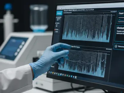
The process of interpreting a standard ultrasound is an incredibly taxing cognitive feat that many people outside the field don’t fully appreciate. A technician must look at flat, 2D images on a screen and, in real-time, mentally arrange those slices into a comprehensive 3D representation of the internal tissue. This creates a significant cognitive burden that often leads to a very long learning curve for students and can occasionally result in scanning inaccuracies even for seasoned pros. When a provider is stuck in this mental bottleneck, they are spending a massive amount of energy just trying to orient themselves spatially, which can take away from the deeper analysis of the pathology they are looking at. It makes the procedure more time-consuming and leaves room for the nagging worry that a small but critical detail might have been missed in the mental reconstruction.
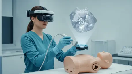
How does the AR-VIU system translate raw ultrasound data into an immersive “X-ray vision” experience for the user?
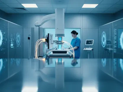
The magic happens when the system streams data into a 3D computer graphics engine known as Unreal Engine, which processes the voxel data to create a high-fidelity 3D representation without any loss of information. This digital model is then projected through an AR/VR headset, allowing the user to see the internal structure superimposed directly over the object’s actual physical location. It feels less like looking at a monitor and more like having true X-ray vision where the skin becomes transparent to the user’s eyes. By simply tilting their head or moving to a different side of the patient, the technician can see different angles of the internal anatomy, making it much more natural to identify and analyze complex structures.
What are the specific hardware innovations that allow this new probe to be more cost-effective and portable than traditional 3D ultrasound systems?
The team at MIT designed a probe that is remarkably compact—it’s actually slightly smaller than a standard deck of cards. Instead of the dense, expensive arrays found in high-end 3D systems, this version uses an ultrasound array configured in the shape of an empty square. This clever geometry, combined with a chirped data acquisition system (cDAQ), allows the device to capture volumetric data using significantly fewer ultrasound elements. Because it uses fewer components, it requires much less power to operate and is far less expensive to build, which opens the door for it to be used in a wider variety of clinical settings beyond just specialized imaging centers.
In the study comparing 18 participants, what did the performance of the novices suggest about the future of medical training?
The study was quite revealing because it showed that when using the AR-VIU system, novices—people who had never even touched an ultrasound before—performed nearly as well as the 9 medical experts in the group. In tasks like identifying hidden objects like screws or springs in gelatin, the 3D visual context provided by the AR headset leveled the playing field almost immediately. For the task of marking “tissue phantoms” to simulate needle placement, the novices found the process intuitive enough to bypass years of traditional 2D training. This suggests that we could drastically accelerate the training process for healthcare providers, moving away from months of learning “mental tomography” and toward a system where the technology handles the spatial orientation for them.
How do the reactions of seasoned experts versus newcomers highlight the potential transition period for this technology in hospitals?
It was interesting to note that while the 9 novices almost universally preferred the AR-VIU system because it felt easier and more intuitive, many of the experts still felt a pull toward the traditional 2D imaging they were accustomed to. This is largely a result of muscle memory and years of professional habit; they have already mastered the difficult task of mental reconstruction, so the 2D screen feels “natural” to them. However, even these experts were quick to point out that the 3D AR system would be an incredible asset for high-stakes tasks like placing a needle for a biopsy or watching the movement of a heart wall. They recognized that while they may not need it for every routine scan, the extra peace of mind and precision it provides during complex procedures is undeniable.
What is your forecast for the integration of real-time 3D ultrasound in standard clinical procedures?
I believe we are heading toward a future where 3D augmented reality becomes the baseline standard for ultrasound, particularly as we further improve the resolution and accuracy of the imaging. Within the next decade, we will likely see these portable, low-cost probes becoming a staple in every emergency room and outpatient clinic, allowing even non-specialists to perform accurate biopsies or rapid cardiac assessments. The “mental tomography” bottleneck will eventually become a relic of the past as the technology matures to a point where the digital and physical worlds are perfectly aligned. This transition will not only make healthcare more accurate by reducing human error but will also make it significantly more accessible by lowering the barrier to entry for performing complex diagnostic tasks.