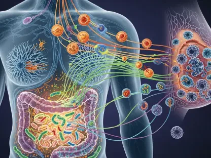
The long-standing struggle to bridge the gap between initial laboratory success and actual therapeutic effectiveness has led the pharmaceutical industry to fundamentally rethink how molecular interactions are measured. For many years, the standard approach involved testing potential drug compounds against isolated proteins in a controlled test tube environment, a method that frequently produced misleading results once those same compounds were introduced into a living system. This fundamental disconnect between artificial laboratory settings and the chaotic, dynamic reality of human physiology has prompted a widespread transition toward live-cell target engagement assays. By observing how a candidate drug binds to its intended protein while that protein remains in its natural habitat, researchers are gaining a level of biological clarity that was once considered unattainable. This transition represents much more than a technical upgrade; it is a conceptual revolution that prioritizes the complex biological context of a disease over the isolated behavior of its individual molecular components. As the industry moves deeper into this era of in-cell validation, the focus is shifting away from simply identifying molecules that can bind to a protein and toward discovering those that can successfully navigate the crowded cellular interior to reach their destination. This strategy provides a much more accurate predictor of how a drug will perform in patients, potentially shortening development timelines and reducing the immense costs associated with late-stage trial failures.
The Limitations: Why Traditional Biochemical Assays Fall Short
The historical reliance on purified proteins for drug screening was largely a matter of convenience and technical limitations, as these isolated systems were easy to handle and allowed for high-throughput testing. This biochemical paradigm served the industry well during the era of simple enzyme inhibitors, providing a straightforward way to rank thousands of chemical compounds based on their affinity for a specific target. However, this simplified model ignored the reality that proteins rarely function in isolation; they are part of a vast, interconnected network of signals and structural supports that define their activity. In a test tube, a protein is stripped of its natural partners, its post-translational modifications, and the specific environment that keeps it in its active shape. Consequently, a drug that appears highly effective in a biochemical assay may fail to work in a cell simply because the protein it is supposed to target does not exist in the same state or configuration as it did during the initial screening process.
Modern drug discovery efforts are increasingly targeting complex multi-protein assemblies and membrane-bound receptors that are notoriously difficult to study outside of a living cell. These targets often require a specific lipid environment or the presence of specific co-factors to maintain the structural integrity needed for drug binding. Furthermore, traditional assays fail to account for the physical barriers a drug must overcome, such as the semi-permeable cell membrane and the various efflux pumps that actively remove foreign substances from the cytoplasm. When researchers rely solely on purified protein data, they often overlook the impact of intracellular drug metabolism, where the cell’s own enzymes might break down a compound before it ever reaches its target. This lack of context explains why so many promising leads in the laboratory fail to show efficacy in the clinic, highlighting the urgent need for screening tools that incorporate the full complexity of a living organism from the very beginning of the research process.
Energy Transfer: Illuminating Interactions in Real Time
To overcome the blind spots of traditional methods, scientists have developed sophisticated systems that utilize light-based energy transfer to detect molecular proximity within intact cells. These techniques, such as Bioluminescence Resonance Energy Transfer, involve the use of a light-emitting donor molecule attached to the target protein and a fluorescent receiver attached to a tracer or the drug itself. When the two molecules come into extremely close proximity—usually within less than ten nanometers—the energy from the donor is transferred to the receiver, creating a distinct light signal that can be measured by sensitive laboratory equipment. This allows researchers to monitor the exact moment a drug molecule interacts with its target in real-time, providing a dynamic view of drug behavior that static biochemical assays simply cannot match. Because the signal is only generated during an actual binding event, it serves as a highly specific indicator of target engagement that is relatively immune to the background noise often found in complex biological samples.
The true power of these bioluminescent tools lies in their ability to function within fully intact, living cells without requiring the destructive processing steps that were common in older methodologies. Historically, measuring drug-target binding often required lysing the cell, a process that inherently disrupts the very environment researchers are trying to study. By keeping the cell alive and healthy, modern assays preserve the natural timing and spatial organization of cellular activities, which is critical for understanding drugs with complex binding kinetics. For instance, some drugs may bind to a target only briefly before being released, while others may form a more permanent bond; these differences can have profound effects on the drug’s ultimate therapeutic impact and its potential for side effects. Being able to observe these interactions as they happen allows for a more nuanced understanding of how a drug’s physical binding translates into a biological response, bridging the gap between molecular chemistry and systemic pharmacology.
Beyond Labels: The Rise of Target-Agnostic Detection
While many engagement assays require specific chemical tracers to function, recent innovations have led to the development of methods that do not rely on pre-existing knowledge of a drug’s binding site. One of the most significant advancements is the use of thermal shift assays adapted for live cells, which take advantage of the fact that proteins become more stable and resistant to heat when they are bound to a drug molecule. By measuring the temperature at which a protein begins to unfold or degrade within a cell, researchers can determine if a candidate compound has successfully reached and stabilized its target. This target-agnostic approach is particularly valuable for exploring the “undruggable” proteome, where many proteins lack well-defined binding pockets or known ligands that could be used as tracers. It allows scientists to screen for interactions across a wide range of proteins simultaneously, turning the target protein itself into a biological sensor that reports on its own status.
This shift toward label-free or agnostic detection significantly expands the scope of what can be achieved in early-stage drug discovery, offering a pathway to treat diseases that have long resisted traditional intervention. For proteins that were previously considered impossible to track, these new assays provide a clear signal of engagement that can be used to guide the optimization of chemical structures. Furthermore, these methods allow researchers to identify unintended interactions where a drug might bind to an off-target protein, potentially causing toxic side effects. By providing a comprehensive view of how a molecule interacts with the entire cellular environment, these technologies help ensure that the most promising and safest candidates are prioritized for further development. This versatility is making live-cell engagement data a cornerstone of modern medicinal chemistry, allowing for the rapid identification of high-quality leads that are more likely to succeed in the rigorous environment of human clinical trials.
Intracellular Geography: Mapping Drug Activity Across Organelles
The modern understanding of cell biology has moved past the simplistic view of the cell as a uniform mixture of chemicals, recognizing instead that it is a highly organized space with distinct functional zones. A drug’s ability to treat a disease often depends not just on whether it enters the cell, but on whether it can reach the specific organelle where the target protein resides, such as the nucleus, the mitochondria, or the endoplasmic reticulum. Each of these compartments has its own unique chemical environment, including different pH levels, ion concentrations, and metabolic enzymes, all of which can influence how well a drug binds to its target. Live-cell assays are now being designed with spatial resolution in mind, allowing researchers to track the engagement of a drug within these specific neighborhoods. This level of detail provides critical insights into why certain drugs might work well in one tissue type but fail in another, as the local microenvironment can either facilitate or hinder the desired molecular interaction.
Understanding these spatial dynamics is essential for developing the next generation of targeted therapies, particularly for diseases like cancer or neurodegenerative disorders where the target proteins are often sequestered in specific parts of the cell. If a drug is highly effective at binding to a protein in the cytoplasm but cannot penetrate the nuclear membrane where the disease-causing activity is actually occurring, it will ultimately fail as a therapeutic agent. By utilizing live-cell imaging and localized sensors, scientists can now visualize the distribution of drug-target interactions across the various compartments of an intact cell. This information allows for the design of “smart” molecules that are specifically engineered to accumulate in the correct organelle, maximizing their efficacy while minimizing the dose needed to achieve a biological effect. Such precision was virtually impossible to achieve using older test-tube methods, which destroy the cell’s architecture and blend all of its components into a single, undifferentiated mixture.
Pipeline Resilience: De-risking Development Through Cellular Insights
Integrating live-cell engagement data early in the drug development process provides a strategic advantage by filtering out compounds that are likely to fail long before they reach expensive clinical trials. This “de-risking” strategy is vital for pharmaceutical companies looking to optimize their research and development budgets and improve the overall success rate of their pipelines. By identifying issues like poor cell membrane permeability or rapid intracellular degradation at the lead optimization stage, researchers can redirect their efforts toward more viable chemical scaffolds. This prevents the “sunk cost” fallacy, where years of effort and millions of dollars are spent on a molecule that was never capable of reaching its target in a living system. The ability to confirm target engagement in a cellular context provides a much higher degree of confidence that the observed biological effects are actually caused by the drug hitting its intended mark, rather than through some secondary or unintended mechanism.
This rigorous focus on cellular reality also helps to clarify the relationship between a drug’s physical binding and its functional outcome, which is often a point of confusion in early discovery. Simply binding to a protein does not always guarantee that the protein’s activity will be inhibited or activated in a way that treats the disease. Live-cell assays allow researchers to correlate the degree of target occupancy with the resulting changes in cellular signaling or gene expression, providing a direct link between chemistry and biology. This mechanistic clarity is particularly important for the development of small-molecule degraders and other novel therapeutic modalities that work through complex, multi-step processes. As these assays become more automated and scalable, they are transitioning from specialized validation tools into the primary method for initial screening. This change ensures that the molecules moving forward in development are those most likely to provide a meaningful benefit to patients, grounded in the reality of human cellular biology.
The adoption of live-cell target engagement assays transformed the landscape of early-stage drug discovery by providing a realistic window into molecular behavior within the living system. These advanced tools allowed researchers to identify and solve permeability and metabolism issues during the initial design phases, significantly reducing the frequency of late-stage failures that previously plagued the pharmaceutical industry. By moving away from isolated biochemical models, the scientific community established a more reliable pathway for translating laboratory discoveries into effective clinical treatments. The insights gained from these cellular studies provided a more robust foundation for clinical trial design, ensuring that dosing and target expectations were based on actual biological performance. Ultimately, this shift encouraged the development of more precise medicines that were better equipped to handle the complexities of the human body, leading to a new era of therapeutic innovation. These advancements demonstrated that prioritizing biological context was the most effective way to ensure that new medicines delivered on their promise to improve patient health.