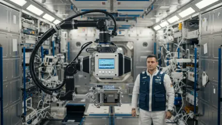

For nearly half a century, the primary method for visualizing internal injuries within the confines of a spacecraft remained restricted to ultrasound, a technique that often falls short when diagnosing complex fractures or identifying serious pulmonary conditions. While ultrasound is an exceptionally versatile tool for soft tissue analysis, its efficacy is heavily dependent on the skill of the operator and the availability of specific acoustic windows in the human body. Recent breakthroughs led by researchers at the Mayo Clinic and the successful execution of the Fram2 mission have finally challenged this status quo by demonstrating that portable digital radiography is not only viable but highly effective in orbit. This milestone represents a fundamental shift in aerospace medicine as human exploration initiatives extend toward the Moon and Mars, where robust and rapid diagnostic capabilities are essential. The transition from sound-based imaging to X-rays provides a much-needed layer of security for crew members.
Technical Adaptation: Portable Radiography in the Orbital Environment
Adapting complex medical hardware for the rigorous environment of space required a departure from the traditional philosophy of building custom-designed, space-rated instruments from scratch. Instead, researchers looked toward commercial off-the-shelf technology that had already been cleared by the FDA for use in terrestrial clinics and emergency departments. By utilizing a compact, energy-efficient digital X-ray generator, engineers were able to overcome the historical hurdles of excessive power consumption and prohibitive bulk that previously prevented radiography from being used on small spacecraft. This lightweight equipment was integrated with a digital flat-panel detector, allowing the system to operate within the extremely limited power budgets of a SpaceX Falcon 9 rocket. This strategy demonstrated that existing high-quality medical technology could be successfully ruggedized to meet the stringent demands of orbital mechanics without requiring decades of development.
One of the most significant barriers to implementing advanced medical diagnostics in space is the requirement for specialized training, which is often difficult for crews to maintain during multi-year missions. The Fram2 mission addressed this challenge by evaluating how quickly non-medical personnel could learn to operate the digital X-ray equipment with high proficiency. In a remarkable display of system usability, three crew members who possessed no formal medical background were able to capture diagnostic-grade images after receiving only four hours of instruction. This ease of operation is a critical factor for missions where every astronaut must serve as a generalist capable of handling various technical and emergency tasks. The ability for non-experts to produce high-resolution radiographs of the chest, abdomen, and limbs ensures that medical diagnostics can be performed accurately and rapidly even when a dedicated radiologist is not physically present on the crew.
Clinical Performance: Validating Diagnostic Quality in Microgravity
Clinical evaluation of the images captured during the mission provided definitive evidence that the diagnostic value of digital radiography is not compromised by the absence of gravity. Independent radiologists conducted blinded assessments, comparing the orbital radiographs to those taken under standard conditions on Earth, and found no statistically significant differences in image clarity or detail. While microgravity did introduce some minor logistical difficulties regarding body positioning—particularly for central-body scans like the pelvis and chest—the resulting images were consistently rated as being of good or near-excellent quality. These results confirmed that the underlying physics of X-ray generation and the sensitivity of digital detectors remain entirely reliable in space. This finding is pivotal because it removes the uncertainty surrounding whether complex diagnostic procedures can provide the same level of accuracy in a deep-space habitat as they do in a modern terrestrial hospital.
Beyond the quality of the diagnostic output, the physical resilience of the hardware was put to the test during the high-vibration phases of launch and the thermal stresses of atmospheric re-entry. Following the return of the equipment to Earth, the X-ray generator and detector underwent a series of rigorous mechanical evaluations to identify any internal degradation or performance shifts. Despite some minor cosmetic wear on the outer casing of the units, the internal components and the stability of the X-ray output were found to be completely unaffected by the journey. This level of durability proved that modern portable medical technology is inherently robust enough to survive the mechanical loads associated with space travel. Establishing that these units can withstand the physical rigors of multiple launches and landings makes them a dependable asset for long-term lunar bases or Martian outposts where equipment failure could have catastrophic consequences for the entire mission.
Operational Resilience: Mechanical Durability and Future Applications
The successful deployment of X-ray technology in orbit offers benefits that extend far beyond human health, introducing a new era of multi-purpose diagnostic utility for space exploration. These portable units provide a dual-use capability that allows crew members to perform non-destructive testing on mission-critical hardware and structural components. For instance, the same generator used to diagnose a suspected fracture can also be used to inspect the internal integrity of complex electronics or the layered fabrics of a pressurized spacesuit. This versatility is indispensable for autonomous missions where spare parts are limited and the ability to detect internal defects before they lead to a failure is a primary safety requirement. By integrating medical imaging with technical maintenance protocols, agencies can maximize the utility of every kilogram of cargo launched into space, ensuring that a single device serves both the biological and mechanical needs of the expedition.
The successful implementation of portable radiography established a new benchmark for medical self-sufficiency during long-duration orbital missions. Medical directors and mission planners prioritized the integration of these digital systems into the standard equipment manifests for upcoming lunar excursions to ensure diagnostic parity with terrestrial clinics. These teams recognized that the ability to synchronize high-resolution X-ray data with Earth-based medical centers facilitated more accurate remote consultations and reduced the risk of diagnostic errors. Furthermore, the decision to adopt dual-use hardware protocols allowed engineers to utilize the same X-ray generators for routine structural integrity checks of critical spacecraft components. By standardizing these imaging procedures, agencies successfully bridged the gap between basic orbital first aid and comprehensive clinical care. This transition proved essential for the safety of crews operating in environments where immediate evacuation to Earth was no longer a viable option.