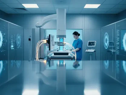

The ability to observe the most intricate biological processes within a living organism represents one of the final frontiers of modern medicine, and today, the Department of Bioengineering at Imperial College London is leading this charge with a series of well-funded initiatives. Recent support from the Wellcome Trust, specifically through the prestigious Bioimaging Technology Development Awards, has catalyzed three flagship projects that aim to decode the complexities of the brain, the metabolic system, and cardiovascular development. By merging high-level engineering with advanced computational science, researchers are pushing past the traditional barriers of medical imaging to observe living systems with unprecedented clarity. These efforts are not merely incremental improvements but are instead transformative leaps that allow for the real-time visualization of cellular behavior. This strategic injection of capital ensures that the next several years, through 2028, will be defined by rapid innovation in how diseases are diagnosed and treated globally.
Breakthroughs in High-Speed Neural Mapping
Visualizing Brain Activity: The Optical Oscilloscope
The project known as the Optical Oscilloscope, spearheaded by Dr. Amanda Foust, addresses a long-standing challenge in neuroscience involving the inability to record rapid electrical signals across three-dimensional brain volumes in real time. Current imaging methods often lack the speed to capture the high-speed communication networks of the brain, but this new initiative utilizes light-field and two-photon microscopy to bridge that critical gap. By synthesizing advanced hardware with deep-learning algorithms, the team can now reconstruct neural signals even when they are obscured by dense, scattering brain tissue. This approach allows for a level of detail that was previously thought impossible without invasive procedures. The system represents a paradigm shift in how neuroscientists approach the study of the central nervous system, providing a dynamic view of electricity in motion. Such capabilities are essential for understanding the fundamental principles of cognition and sensory processing in various complex organisms.
This technology utilizes high-performance GPUs and FPGAs to process massive datasets instantaneously, moving beyond the physical speed limitations associated with traditional mechanical scanning methods. By removing the mechanical constraints that once slowed down data acquisition, researchers can now monitor thousands of neurons simultaneously without sacrificing the temporal resolution required to see individual action potentials. Beyond its technical merits, the project has immediate clinical implications, specifically regarding the ongoing study of Alzheimer’s disease and other degenerative conditions. By investigating neural hyperexcitability and disrupted neurovascular coupling, the Optical Oscilloscope is poised to become a vital tool for developing therapies for a wide range of neurological conditions linked to electrical dysfunction. The project emphasizes a move toward holistic data collection, where the interactions between different brain regions are viewed as a single, unified system rather than isolated fragments.
Clinical Applications: Deciphering Complex Neuropathology
The practical utility of high-speed neural mapping extends far beyond the laboratory, offering a new lens through which researchers can examine the progression of debilitating brain disorders. In the context of Alzheimer’s research, the ability to see how electrical signals misfire in real time provides a direct link between cellular abnormalities and observable cognitive decline. This level of insight allows for the identification of early-stage biomarkers that were previously invisible to conventional MRI or CT scans. Furthermore, the integration of machine learning allows for the prediction of disease trajectories, enabling personalized treatment plans tailored to the specific electrical profile of a patient. This predictive capability is a cornerstone of the shift toward precision medicine, where interventions are timed to maximize efficacy. As these tools become more refined, the gap between bench research and bedside application continues to shrink, promising faster transitions for experimental therapies.
Moreover, the versatility of the Optical Oscilloscope allows it to be adapted for various other conditions, such as epilepsy or traumatic brain injury, where timing is everything. Understanding the exact sequence of events during a seizure or the immediate aftermath of a concussion can lead to more effective emergency interventions. The research team is currently focused on optimizing the signal-to-noise ratio in deeper brain structures, which have traditionally been the hardest to image due to light scattering. By overcoming these optical hurdles, the project ensures that no part of the brain remains in the dark. The collaborative nature of this work also means that the software and hardware blueprints are shared with the broader scientific community, fostering a global environment of innovation. This open-source approach accelerates the pace of discovery, ensuring that the benefits of bioimaging are felt across different medical disciplines and geographical borders during the current research cycle.
Innovations in Non-Invasive Deep-Tissue Monitoring
Advancing Super-Resolution Ultrasound: Imaging Micro-Circulation
Led by Professor Mengxing Tang, the second major initiative focuses on dynamic 3D super-resolution ultrasound to peer into the body’s micro-circulation without the need for invasive surgery. Historically, viewing the smallest blood vessels deep within a living organism at high resolution has been nearly impossible due to the limitations of wave physics. This project overcomes that hurdle by combining ultrafast volumetric ultrasound with genetically encoded acoustic reporters, which essentially turn specific cells into detectable signal emitters for ultrasound waves. This allows for the visualization of blood flow at the capillary level, providing insights into how tissues are oxygenated and how nutrients are distributed throughout the body. The resolution achieved by this system is an order of magnitude higher than standard clinical ultrasound, making it a powerful tool for early disease detection. It offers a clear view of the structural changes that precede major health crises.
The primary focus of this specific research is metabolism, particularly tracking how insulin-producing cells in the pancreas behave and how gut cells communicate with the rest of the body. By monitoring these interactions in a non-invasive manner, researchers can gain a deeper understanding of the mechanisms behind type 2 diabetes and metabolic syndrome. The use of acoustic reporters means that specific cell populations can be highlighted without the use of radioactive tracers or toxic contrast agents. This makes the technology safer for long-term monitoring and repeatable studies, which is essential for tracking the efficacy of new drug treatments. Furthermore, the ability to see the metabolic state of an organ in real time provides a much more accurate picture than blood tests alone. This granular data allows for a more nuanced understanding of how lifestyle factors and genetics interact to influence metabolic health, paving the way for highly targeted nutritional and medical interventions.
Bacterial Engineering: Redefining Metabolic Diagnostics
A groundbreaking aspect of this ultrasound work involves engineering gut bacteria to be visible via ultrasound, potentially allowing for the diagnosis of infections or inflammation through routine scans. This innovative approach utilizes the natural presence of bacteria in the human microbiome to act as internal sensors that report on the health of the digestive tract. By modifying these microbes to produce small gas vesicles, they become highly reflective to ultrasound waves, allowing their location and density to be mapped precisely. This shift toward non-invasive monitoring could eventually replace painful or expensive endoscopic procedures, making diagnostic care more accessible and patient-friendly for millions. The ability to “see” the microbiome in action opens up new possibilities for treating inflammatory bowel disease and other chronic gut conditions. It represents a significant step forward in the integration of synthetic biology with traditional medical imaging.
Beyond simple diagnostics, these engineered bacteria could also be used to deliver localized therapies exactly where they are needed most. For instance, bacteria could be programmed to release anti-inflammatory compounds only when they detect a specific chemical marker of disease, with ultrasound used to confirm their arrival at the target site. This dual-purpose role as both a diagnostic tool and a delivery vehicle highlights the multidisciplinary nature of modern bioengineering. The reduction in patient discomfort and the lowering of procedural costs are major drivers for the adoption of this technology in clinical settings. As the research progresses through 2027, the team expects to refine the sensitivity of these bacterial sensors, allowing for the detection of even the most subtle changes in the gut environment. This proactive approach to health monitoring shifts the focus from treating advanced symptoms to managing the earliest signs of biological imbalance, significantly improving patient outcomes.
Mapping the Mechanical Forces of Life
Tracking Cardiovascular Development: Insights from Live Models
Imperial College London is also playing a critical role in a collaborative effort to study the mechanical forces that guide heart formation during the earliest stages of life. Led by Professor Julien Vermot at Imperial, this project utilizes the natural transparency of live zebrafish larvae to create detailed 3D maps of physical stresses and strains during cardiac development. This study aims to provide a comprehensive resource for the global research community by documenting the entire timeline of heart morphogenesis with high precision. Understanding how physical forces like fluid shear stress and tissue tension shape the growing heart is vital for identifying the root causes of congenital defects. The use of zebrafish is particularly advantageous because their hearts develop rapidly and share many genetic similarities with the human heart. This allows researchers to observe developmental processes that are usually hidden deep within the womb.
By utilizing high-speed confocal microscopy and specialized fluorescent markers, the team can track individual cells as they migrate and differentiate to form the complex architecture of the heart. This research has revealed that mechanical signals are just as important as genetic instructions in determining the final shape and function of the organ. If these forces are imbalanced, it can lead to structural abnormalities that persist throughout a person’s life. The data generated from these observations is being used to create sophisticated computer models that can simulate heart development under various conditions. These models serve as a virtual laboratory where scientists can test how different mutations or environmental factors affect cardiac health. The project emphasizes the importance of biomechanics in the broader field of developmental biology, proving that life is shaped by physics as much as it is by chemistry. This knowledge is fundamental for the future of regenerative medicine.
Future Perspectives: Integrating Multidisciplinary Engineering
The synthesis of data from multiple disciplines, including machine learning, genetic engineering, and optics, highlights a broader trend in the evolution of bioengineering. The shift from static 2D images to real-time, volumetric data ensures that the next generation of medical research will be defined by a holistic, non-invasive understanding of the body. This integration allows for a more comprehensive view of how different systems, such as the cardiovascular and nervous systems, interact with one another to maintain homeostasis. For example, understanding how blood flow in the brain influences neural activity is a key area of interest that benefits from the combination of ultrasound and optical techniques. This interdisciplinary approach breaks down the silos that have traditionally separated different fields of medicine, fostering a more collaborative and efficient research environment. It is this synergy that drives the most significant breakthroughs in modern healthcare.
Furthermore, the development of these advanced imaging platforms creates a robust infrastructure for future scientific exploration. The tools being built today at Imperial will serve as the foundation for countless studies in the years to come, ranging from cancer research to tissue engineering. As imaging resolution continues to improve, the ability to see the molecular interactions that drive life will become a routine part of medical practice. This will likely lead to the discovery of new biological pathways and the development of more effective, less toxic drugs. The commitment to non-invasive techniques also reflects a growing emphasis on patient-centered care, where the goal is to provide high-quality data without the risks associated with surgery. By continuing to push the boundaries of what is possible, the Department of Bioengineering is ensuring that the future of medicine is brighter, clearer, and more accessible for everyone, regardless of the complexity of their condition.
Advancing the Precision of Human Healthcare
The strategic initiatives at Imperial College London successfully demonstrated that the future of medical diagnostics rests on the seamless integration of physical engineering and biological insight. By securing the Wellcome Trust funding, the research teams established a new framework for observing the human body that bypassed the limitations of traditional, invasive techniques. The development of the Optical Oscilloscope and super-resolution ultrasound provided clinicians with the tools necessary to visualize disease progression at its most fundamental level. These projects moved the needle toward a more proactive form of medicine, where the focus transitioned from reactive treatment to early, high-precision detection. The collaborative efforts involving cardiovascular mapping further solidified the role of mechanical physics in understanding life, offering a more complete picture of human development than had ever been recorded before.
Ultimately, the advancements made during this period provided a clear roadmap for the next steps in clinical implementation and technological refinement. Medical professionals were encouraged to adopt these non-invasive platforms to reduce patient risk and improve the accuracy of longitudinal health monitoring. The integration of artificial intelligence into bioimaging was finalized as a standard requirement for managing high-throughput data, ensuring that no vital signal was lost in the noise of biological complexity. As these technologies moved toward widespread commercial availability, they offered a practical solution for addressing the rising global burden of chronic diseases. The success of these initiatives proved that when diverse scientific disciplines converged, the resulting innovations possessed the power to redefine the boundaries of what was once considered medically possible.