
The intricate dance of life within a human cell relies on more than just the genetic blueprints stored in the nucleus or the proteins that perform the heavy lifting. For decades, biology textbooks have described the cell membrane as a passive container—a structural scaffold that simply holds proteins in place like a wall holds windows. However, recent findings from chemists at the Massachusetts Institute of Technology have effectively dismantled this “scaffold dogma.” Their research proves that the lipid bilayer is far from a neutral bystander; it is a primary driver of cellular behavior. By altering its own chemical environment, the membrane can seize control of vital signaling pathways, dictating when a cell stays dormant and when it begins the rapid, uncontrolled division that defines cancer.
This paradigm shift suggests that the physical housing of a cell possesses an instructional capacity previously reserved for DNA and complex enzymes. By focusing on the lipid environment, scientists are uncovering a hidden layer of regulation that could explain why certain aggressive tumors are so resilient. Understanding how the membrane communicates with internal proteins provides a missing link in the study of malignancy, moving beyond the protein-centric view of disease.
Beyond the Scaffold: A New Era in Cellular Biology
The transition away from viewing the membrane as a mere boundary marks a significant turning point in molecular biology. Historically, the lipid bilayer was seen as a fluid but inert stage where the “real” work was performed by membrane-bound proteins. This perspective limited the scope of cancer research, as investigators focused almost exclusively on mutations within the proteins themselves. The recent work published in the journal eLife reveals that the stage itself can influence the performance of the actors. By changing the ratio of specific lipid molecules, the membrane exerts physical and chemical forces that can fundamentally alter protein function.
Moreover, this research indicates that the membrane acts as a sophisticated signaling hub rather than a static barrier. It possesses the ability to organize proteins into clusters, concentrate signaling molecules, and even change its own curvature to facilitate specific biological processes. When this organizational capacity is hijacked by cancer cells, the result is a breakdown in the normal communication channels that prevent overgrowth. Consequently, the lipid environment is now recognized as a potent regulator of the cellular “on-off” switches that govern life and death.
The EGFR Connection: A High-Stakes Molecular Switch
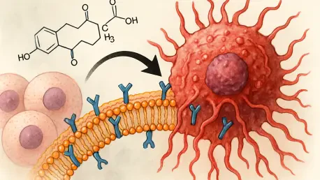
At the heart of this discovery is the Epidermal Growth Factor Receptor (EGFR), a protein that acts as a gatekeeper for cell growth and tissue repair. In a healthy system, EGFR functions like a precision-engineered switch, activating only when it receives a specific “go” signal from an external growth factor. This activation allows the body to heal wounds or replace old cells in a controlled manner. However, in aggressive malignancies such as glioblastoma and lung cancer, this regulatory process fails, leading to the relentless proliferation of tumor cells.
While scientists previously blamed an overabundance of the EGFR protein for these failures, the study reveals a more subtle culprit: the surrounding lipids can force these switches into a permanent “on” position. This shift occurs even in the absence of traditional growth signals, providing a chemical loophole that cancer cells exploit to thrive. The discovery that the lipid environment can trigger EGFR independently of external ligands suggests that current treatments targeting only the ligand-binding site may be fundamentally incomplete.
Decoding the Impact of Negative Charge and Membrane Rigidity
The behavior of the EGFR switch is dictated by two primary chemical factors within the membrane: electrical charge and physical stiffness. In healthy cells, negatively charged lipids make up about 15% of the membrane, allowing the receptor to function normally within its regulatory bounds. When that concentration spikes to 60%—a level often observed in tumor cells—the receptor undergoes a structural change that locks it into an active state. This high-charge environment essentially “tricks” the cell into a state of constant growth by pulling the receptor into a conformation that mimics the presence of a growth signal.
Conversely, the presence of cholesterol adds rigidity to the membrane, acting as a physical brake. This stiffness makes it harder for the receptor to change shape, preventing it from sending growth signals even when charged lipids are present. This delicate balance between the “push” of negative charge and the “pull” of rigidity represents a critical regulatory mechanism in cancer progression. Tumors appear to manipulate this balance, favoring a more fluid and highly charged environment to maintain their rapid expansion and survival under stress.
Probing the Molecular Machinery: Insights from the Schlau-Cohen Lab
To uncover these mechanics, Professor Gabriela Schlau-Cohen and lead author Shwetha Srinivasan utilized a combination of cutting-edge synthetic biology and high-resolution imaging. Because membrane proteins are notoriously difficult to study in isolation due to their requirement for a lipid environment, the team developed “nanodiscs.” These are synthetic models that mimic the natural cellular environment, allowing for the precise control of lipid composition. This innovation allowed the researchers to isolate the effects of specific lipids on the EGFR protein without the noise of a full living cell.
By employing single-molecule Fluorescence Resonance Energy Transfer (FRET), they attached fluorescent tags to the EGFR protein to measure nanometer-scale movements in real-time. These observations, supported by atomic-level molecular dynamics simulations, provided the first clear look at how lipid chemistry physically twists and pulls the receptor into a pro-cancer conformation. This level of detail offered insights that traditional clinical observations could never reach, proving that the chemical makeup of the membrane directly modulates the physical shape of signaling proteins.
From Biology to Therapy: Strategies for Targeting the Lipid Environment
The realization that membrane chemistry drives cancer growth offered a promising new framework for oncological treatment that looked beyond the genetic sequence. Many patients developed resistance to drugs that targeted the EGFR protein directly because the cell eventually bypassed the inhibited site through mutation. A more resilient strategy involved “remodeling” the membrane environment rather than the protein itself, effectively changing the soil in which the cancer grew. Potential therapeutic avenues included developing agents that neutralized excessive negative charges on the surface of tumor cells or adjusted membrane fluidity to suppress overactive signaling.
By shifting the focus from the “engine” (the protein) to the “fuel” (the lipids), researchers paved the way for a new generation of therapies. These future interventions targeted the biophysical properties of the cell, making it much harder for tumors to adapt through simple genetic changes. Scientists explored the possibility of using lipid-modifying enzymes to restore the healthy 15% charge balance, thereby forcing overactive receptors back into a dormant state. This approach not only offered a way to overcome drug resistance but also suggested a method for stabilizing cellular health across a wide range of different cancer types.


