
The detection of rhythmic internal contractions during a real-time ultrasound scan provides a pathognomonic sign that confirms the presence of a living organism. As international travel continues to rise in 2026, medical practitioners in temperate regions are increasingly encountering tropical conditions that were once considered rare or exotic. Among these, cutaneous myiasis stands out as a particularly distressing condition for patients, involving the infestation of living human tissue by dipterous fly larvae. The most frequent culprit in the Americas is the human botfly, Dermatobia hominis, which utilizes a unique biological strategy to develop within the host’s subdermal layers. While the visual symptoms of this infestation often mimic common bacterial infections, the precision of modern high-frequency ultrasound allows for an immediate and accurate diagnosis, bypassing the guesswork that previously led to ineffective treatments. This transition toward real-time imaging represents a significant shift in travel medicine, where the ability to see beneath the skin’s surface transforms a complex parasitological puzzle into a manageable clinical case.
Overcoming the Clinical Mimicry of Botfly Infestations
The Diagnostic Challenge: Identifying Parasitic Nodules
Cutaneous myiasis typically presents as a firm, erythematous, dome-shaped nodule that bears a striking resemblance to a standard bacterial boil or a furuncle. This clinical mimicry is the primary reason why many patients undergo weeks of unnecessary antibiotic therapy before the true cause of their discomfort is identified. The larva creates a small central respiratory pore in the host’s skin, which is essential for its survival but can be easily mistaken for the “head” of a maturing abscess. Because the lesion is often painful, tender, and associated with localized swelling, a physician might naturally gravitate toward a diagnosis of cellulitis or a deep-seated infection. However, the underlying pathology is entirely different; while an abscess is a collection of necrotic tissue and white blood cells, a botfly infestation is a dynamic biological process involving a living, breathing organism that is actively feeding and growing within the subcutaneous space.
The danger of misdiagnosing these nodules as simple infections extends beyond mere treatment delays. If a clinician, suspecting a standard abscess, attempts a traditional incision and drainage procedure, the results can be catastrophic for the patient’s recovery. Standard surgical tools are not designed to remove an intact, clinging larva, and the pressure applied during a drainage attempt can easily rupture the parasite’s body. A ruptured larva releases foreign proteins and biological material directly into the host’s bloodstream and surrounding tissue, which frequently triggers a massive inflammatory response. This can lead to secondary bacterial infections that are far more difficult to manage than the initial infestation. Furthermore, leaving fragments of the exoskeleton behind can cause long-term granulomatous reactions, making it imperative that the diagnosis is confirmed through non-invasive imaging before any surgical intervention is considered.
Contextual History: Moving Beyond Surface Observations
The path to an accurate diagnosis begins with a thorough exploration of the patient’s recent travel history, particularly if they have visited rural or forested areas in Central or South America. In these regions, the life cycle of the botfly is a common biological occurrence, yet for a traveler returning to a major metropolitan area, the symptoms may feel entirely alien. The biological mechanism is fascinating and complex; the adult fly often captures a mosquito or another blood-sucking insect to carry its eggs to a human host. When the carrier insect feeds, the warmth of the host’s skin triggers the eggs to hatch, and the tiny larvae immediately burrow into the skin. This history is often the first clue that a persistent skin lesion is not a simple staph infection but a “living passenger” acquired during a tropical excursion. Relying only on visual inspection without considering these geographical factors often leads to a cycle of failed medical interventions and increasing patient anxiety.
Modern medicine in 2026 emphasizes the integration of patient narrative with advanced diagnostics to avoid the pitfalls of surface-level observations. When a patient reports a sensation of something moving or “crawling” beneath the skin, or describes intermittent, sharp, stabbing pains, these are classic indicators of larval activity that should prompt immediate imaging. These subjective reports are frequently dismissed by clinicians who are more accustomed to treating typical skin flora infections, yet they are the hallmark of a parasitic infestation. By recognizing that the botfly larva is a physical entity that requires oxygen and space to grow, doctors can shift their focus from systemic antibiotics to localized imaging. This transition allows for the identification of the subdermal cavity where the larva resides, providing a definitive answer that visual inspection and palpation simply cannot offer, especially in the early stages of the infestation.
The Technical Advantages of High-Frequency Ultrasound
Structural Analysis: Identifying Solid Entities Through Imaging
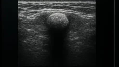
In the modern radiology suite, the use of high-frequency linear transducers—specifically those operating at 13-MHz or higher—has revolutionized the way subcutaneous lesions are evaluated. When a patient presents with a suspected botfly infestation, the initial gray-scale imaging provides the first definitive evidence of a solid structure. In a typical abscess, the ultrasound screen would display a hypoechoic or dark fluid collection, representing the liquid pus and debris. In contrast, a botfly larva appears as a distinct, echogenic mass within the tissue. The resolution provided by high-frequency probes is so refined that it can often visualize the segments of the larva’s body, allowing the radiologist to determine the exact size and orientation of the parasite. This clarity is essential for distinguishing between an inflammatory fluid pocket and a physical, organized biological organism.
One of the most reliable indicators found during these scans is known as posterior acoustic shadowing. This phenomenon occurs when the ultrasound waves encounter a material that is significantly denser than the surrounding human tissue, such as the chitinous exoskeleton of a fly larva. As the sound waves hit this dense surface, they are either absorbed or reflected so strongly that a dark “shadow” is cast directly behind the object on the resulting image. This finding is a game-changer for the medical team, as it confirms they are looking at a solid physical entity rather than a liquid infection. While a foreign body like a wood splinter might also produce shadowing, the specific shape and location of the shadowing in relation to the echogenic mass help the radiologist narrow down the possibilities. This technical precision ensures that the clinical team can approach the treatment phase with a complete understanding of what lies beneath the surface.
Real-Time Assessment: Capturing the Movement of Living Parasites
The true power of ultrasound in diagnosing myiasis lies in its ability to perform a dynamic, real-time assessment of the affected area. Unlike a static X-ray or a CT scan, which provides only a single moment in time, a live ultrasound allows the clinician to observe the lesion for several minutes. During this period, the “wriggle” factor becomes the most important diagnostic clue. The human botfly larva is not a dormant object; it is a muscular organism that performs rhythmic, peristalsis-like contractions to move within its burrow and maintain its position. Witnessing these internal movements on the ultrasound monitor is a pathognomonic sign, meaning it is a finding so specific to a living organism that it effectively rules out all other non-living mimics. This mechanical shifting is unmistakable once observed, providing a level of diagnostic certainty that is rare in clinical practice.
The importance of this dynamic observation cannot be overstated when differentiating a living parasite from a static foreign body or a vascularized tumor. While a tumor might appear as a solid mass and even show some internal patterns, it will never exhibit the rhythmic, mechanical contractions of a larva. Similarly, a piece of glass or a thorn lodged in the skin will remain perfectly still regardless of how long it is observed. The ability to see the larva move in response to the pressure of the ultrasound probe or simply through its own biological cycles provides the clinician with an immediate confirmation of life. This real-time feedback is invaluable for both the doctor and the patient, as it replaces suspicion with visual proof. In the fast-paced environment of an emergency department or a travel clinic, this rapid confirmation allows for the immediate initiation of extraction protocols, saving the patient from days of uncertainty.
Differentiating Myiasis From Other Skin Conditions
Logical Framework: Comparing Ultrasound Signatures
To provide the most accurate care, clinicians must synthesize various ultrasound signatures to rule out several conditions that frequently present with similar symptoms. The diagnostic process is essentially a process of elimination based on specific imaging characteristics. For instance, a vascular tumor may appear as a solid, echogenic mass similar to a larva, but a tumor will typically show internal blood flow patterns on a Doppler scan rather than the mechanical, muscular contractions of a parasite. On the other hand, a foreign body such as a splinter will show posterior shadowing and significant surrounding inflammation, but it will lack both internal blood flow and any form of spontaneous movement. By methodically checking for these four key traits—echogenicity, shadowing, movement, and internal flow—the radiologist can pinpoint the exact nature of the lesion with high confidence.
The living larva is unique because it is the only subcutaneous structure that combines an echogenic body and posterior shadowing with spontaneous internal movement and a specific type of inflammatory rim. When these elements are viewed together, the “signature” of the botfly becomes undeniable. Modern imaging protocols in 2026 have streamlined this process, allowing for a rapid “yes or no” determination in the radiology suite. This systematic approach prevents the patient from being subjected to more invasive and expensive tests, such as an MRI, which would be unnecessary once the ultrasound has provided a definitive answer. By establishing a clear logical framework for interpreting these scans, the medical community has improved the speed and accuracy of parasitic diagnosis, ensuring that travel-acquired conditions are handled with the same precision as more common domestic ailments.
Vascular Analysis: The Role of Color Doppler in Detection
Color Doppler imaging serves as a vital secondary tool in the diagnostic process, offering insights into the host’s inflammatory response. When the Doppler mode is activated, it detects the velocity and direction of blood flow in the tissue surrounding the suspected larva. In cases of myiasis, there is almost always a significant amount of peripheral hyperemia—an intense increase in blood flow to the area as the host’s immune system attempts to respond to the parasite. This “vascular signature” explains the clinical symptoms of heat, redness, and throbbing pain that the patient experiences. By visualizing this intense ring of inflammation, the clinician can better understand the extent of the tissue involvement and the severity of the host’s reaction. This information is critical for determining whether the patient might also require anti-inflammatory medication or if the infestation has caused a significant localized vascular response.
In addition to confirming inflammation, Color Doppler helps to distinguish the larva from highly vascularized soft-tissue tumors. While the larva itself will not show internal blood flow signals—since its internal systems do not mirror human vascularity—the tissue immediately adjacent to its burrow will be highly active. In contrast, a hemangioma or other vascular tumor would show blood flow throughout the mass itself. This distinction is vital for planning any subsequent intervention. Knowing that the blood flow is restricted to the periphery allows the surgeon to proceed with extraction knowing that the central mass is a parasite and not a blood-filled growth. This level of detail ensures that the treatment plan is tailored to the specific biological reality of the infestation, minimizing the risk of heavy bleeding or other complications during the removal process.
Improving Patient Outcomes and Treatment Precision
Surgical Planning: Mapping for Safe Extraction
Once the presence of a larva was confirmed through imaging, the focus shifted toward the safest possible removal. Having a precise ultrasound map of the larva’s size, depth, and orientation was essential for a successful recovery in recent clinical cases. Imaging allowed the medical professional to plan the extraction with surgical precision, identifying the exact angle at which the larva was lodged. This was particularly important for using “suffocation” techniques, where the respiratory pore is covered with a substance like petrolatum to force the larva toward the surface. By knowing the larva’s orientation, the doctor could anticipate its movement and be ready to grasp it with forceps the moment it emerged to breathe. This preemptive knowledge reduced the total time of the procedure and significantly lowered the stress levels for the patient.
Furthermore, ultrasound proved invaluable for identifying “multiple infestations,” which can occur when a traveler is exposed to a high density of carrier insects. In several recorded instances, a patient presented with one obvious nodule, only for the ultrasound to reveal two or three additional larvae burrowed in the same general area that had not yet created a visible pore. Without the use of a wide-area ultrasound scan, these secondary parasites would have been missed, leading to a recurrence of symptoms just days after the initial treatment. The ability to visualize the depth also ensured that no fragments were left behind after the procedure. Because the barbs on a botfly larva can make extraction difficult, the post-procedural scan became a standard step to verify that the subdermal cavity was entirely clear, ensuring the wound could heal without further complication or granulomatous inflammation.
Strategic Medicine: Addressing Global Trends in Travel Medicine
As global mobility remained a defining characteristic of society in 2026, the appearance of “travel-acquired” myiasis in non-endemic regions became a practical reality for emergency departments worldwide. Physicians in cities far from the tropics found that they could no longer rely solely on their previous clinical experience, as parasites like Dermatobia hominis crossed borders with their hosts. The adoption of high-frequency ultrasound as a first-line diagnostic tool served as an accessible and inexpensive bridge, allowing doctors to solve complex parasitological puzzles with equipment already available in most clinics. The study of these cases demonstrated that a high index of suspicion, combined with a simple probe placement, could prevent weeks of unnecessary antibiotic use. This approach emphasized a fundamental principle: that the most effective diagnostic tools are those that allow for the direct observation of biological behavior in real time.
The insights gained from these diagnostic advancements offered a clear roadmap for future travel medicine protocols. Clinicians were encouraged to prioritize non-invasive imaging over exploratory surgery, recognizing that the “wriggling signature” of a larva provided more information than a physical exam ever could. By opting for ultrasound with Color Doppler early in the clinical pathway, medical teams provided faster, more accurate, and significantly less traumatic care for their patients. The final takeaway from these developments was one of clinical patience and technological integration. By simply waiting to see if a mysterious lump moved under the probe, doctors avoided the risks of misdiagnosis and ensured that patients returned to health with minimal intervention. This evolution in practice underscored the importance of maintaining a global perspective in medicine, where the tools of the modern radiology suite are used to meet the challenges of an increasingly interconnected world.