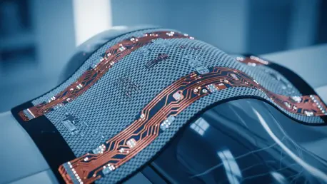

Radiology departments have long struggled with the paradox of patient comfort versus image resolution, where even the slightest movement can ruin a high-field scan and lead to diagnostic uncertainty. The ongoing shift in 2026 toward flexible radiofrequency coils marks a significant departure from the rigid, cumbersome arrays that dominated the previous decade of medical imaging. These flexible designs conform tightly to the unique contours of a patient’s body. This physical proximity brings the receiving elements closer to the target tissue, which significantly improves the signal-to-noise ratio. However, maintaining this quality during inevitable patient movement requires more than just a soft exterior; it demands sophisticated engineering that can compensate for shifting electromagnetic fields. As researchers refine these stretchable materials, the focus has turned to how these coils can maintain their electrical properties while being pulled, bent, or compressed during the dynamic environment of a live clinical examination.
Material Resilience: Ensuring Conductivity Under Stress
Stretchable Conductors: The Science of Elastic Circuitry
One of the most effective methods for maintaining signal integrity during motion involves the use of specialized conductive polymers and liquid metal alloys that exhibit constant impedance despite physical deformation. Conventional copper traces often suffer from micro-fractures or significant resistance changes when repeatedly flexed, but these modern materials allow for high elasticity without sacrificing electrical conductivity. By integrating these stretchable conductors into a flexible substrate, engineers have created coils that maintain a stable resonance frequency even when a patient expands their chest or adjusts a limb. This stability is crucial because any change in the coil’s shape can detune the circuit, causing a drop in the signal-to-noise ratio that manifests as graininess or artifacts in the final image. These resilient materials act as the first line of defense, ensuring that the physical act of stretching does not translate into a loss of critical diagnostic data.
Impedance Control: Maintaining Resonance During Flexion
Beyond material flexibility, the arrangement of individual coil elements plays a vital role in mitigating the effects of motion by utilizing advanced overlapping patterns and decoupling circuits. In a flexible array, the distance between neighboring elements fluctuates as the coil bends, which can lead to unwanted magnetic coupling and signal interference between channels. To counter this, developers have implemented active decoupling strategies that dynamically adjust the electrical isolation between elements in real time. This ensures that each sensor in the array operates independently, capturing a precise slice of data without being influenced by the movement of adjacent sections. When combined with high-density designs that feature smaller, more numerous elements, these arrays can maintain a high degree of sensitivity across the entire surface area. This distributed sensing approach allows the system to recover lost information by oversampling the region of interest.
Dynamic Compensation: Technological Integration for Stability
Sensor Embedding: Synchronizing Motion and Signal
Real-time motion tracking has emerged as a transformative solution, where miniature sensors are embedded directly into the flexible fabric of the MRI coil to monitor displacement as it happens. These sensors, often utilizing optical or ultra-low-power radio signals, transmit the precise coordinates of the coil’s position to the scanner’s central processing unit. This allows the gradient fields and pulse sequences to be adjusted on the fly, effectively following the patient’s movement to maintain a consistent frame of reference throughout the scan. For instance, if a patient’s breathing causes a three-millimeter vertical shift, the system can recalibrate the imaging plane in milliseconds to compensate for that change. This level of synchronization ensures that the image remains sharp, even if the physical coil is constantly undulating. By turning the coil into an active data-gathering tool, clinics have significantly reduced the frequency of motion-induced artifacts.
Algorithmic Refinement: The Final Layer of Image Clarity
To maximize the performance of these adaptive systems, healthcare facilities moved toward integrating AI-driven reconstruction algorithms that could intelligently fill in data gaps caused by sudden or erratic patient shifts. These software solutions were trained on vast datasets of motion patterns, allowing them to distinguish between genuine anatomical features and motion-related distortions with high precision. In practice, the adoption of flexible coils was complemented by rigorous calibration protocols that ensured each array was optimized for specific body types and motion profiles. Radiology teams successfully implemented these tools by prioritizing hardware that offered both high geometric flexibility and robust electronic shielding. Ultimately, the industry shifted its focus toward a holistic approach where material science and digital compensation worked in tandem. This progression ensured that clinicians obtained laboratory-quality images, making the scanning process faster and more reliable.