
The inherent swarming motility of bacterial colonies provides a unique medium for storing information that can be analyzed using standard optical scanning rather than genomic sequencing. This technological leap, spearheaded by researchers at Columbia University, represents a shift from observing natural microbial behaviors to utilizing them as specialized hardware. For years, the scientific community has sought ways to bridge the gap between biological systems and digital data storage. While DNA-based storage has made strides, its reliance on expensive and time-consuming sequencing remains a bottleneck. By engineering Escherichia coli to manifest environmental history as macroscopic patterns, scientists have created a “living recorder” that is both accessible and scalable. This approach leverages the collective intelligence of millions of bacteria, turning their movement into a readable language that describes their surroundings in vivid detail, from chemical exposures to light intensity. This breakthrough marks the beginning of an era where biology and data science merge into a single, visible platform for monitoring the world.
Harnessing Natural Locomotion: Swarming as a Data Medium
The Biological Canvas: Mechanisms of Collective Movement
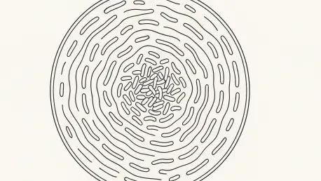
Swarming motility is a coordinated collective behavior where bacteria undergo radical physical transformations to move rapidly across semi-solid surfaces like agar. During this process, individual cells elongate and grow numerous flagella, allowing them to travel in rhythmic waves that expand outward from a central point. This travel is more than mere locomotion; it is an emergent property that integrates cellular signals over time and space to create a physical history of the growth environment. By selecting a specific hypermotile strain of E. coli known as MG1655hm, the researchers ensured that the colony expansion was both rapid and highly reproducible, providing a stable canvas for recording data. The predictability of this expansion is crucial, as it allows any deviation in growth to be attributed to environmental factors. As the bacteria communicate and move together, they leave behind a structural trail that reflects the chemical and physical conditions they encountered during their journey.
To transform this natural expansion into a functional data storage system, the researchers introduced high-copy plasmids containing inducible promoters that control motility-related genes. This setup effectively “hijacks” the colony’s expansion mechanism, linking the rate and style of growth to the presence of external stimuli such as specific chemicals or light. As the bacteria move outward from their point of origin, the physical shape, density, and texture of the resulting colony serve as a permanent, visible record of the conditions encountered during the growth phase. This method of information capture is unique because it records data dynamically as the organism interacts with its environment, rather than requiring a post-growth synthesis of DNA. The final result is a macroscale pattern that can be seen by the naked eye, representing hours or days of environmental exposure integrated into a single, cohesive image. This development paves the way for biological sensors that are as easy to read as a photograph, yet maintain scientific precision.
Strategic Selection: Genetic Pens for Information Encoding
The precision of this living recording device depends on the strategic selection of genes within the bacterial motility hierarchy. The research team identified four primary candidates—lrp, fliA, rpoS, and cheZ—to act as the genetic “pens” that draw the patterns on the agar. Each of these genes occupies a specific level in the regulatory network of E. coli, governing different aspects of how the bacteria move and respond to their environment. For instance, lrp sits at the top of the hierarchy and activates the master regulator of flagellar biosynthesis, making it a powerful switch for starting or stopping movement. Meanwhile, fliA functions as a flagellum-specific sigma factor that controls the expression of later-stage genes required for motility. By targeting these specific components, the researchers could fine-tune the bacterial response to produce distinct visual outputs. This level of control allows for the creation of various “inks” that respond to different triggers, making the system modular.
Beyond simple movement, the selection of genes like rpoS and cheZ provided the ability to manipulate the intricate textures and density of the colonies. The rpoS gene, typically associated with the general stress response in E. coli, acts as an indirect repressor of motility, which the team used to create compact and highly detailed patterns. Meanwhile, cheZ controls the chemotactic signaling that determines the length of forward “runs,” allowing for fine-tuned control over the jaggedness and expansion radius of the colony. This sophisticated manipulation of the internal genetic hierarchy ensures that every visual aspect of the pattern—from its diameter to its internal ring structure—carries specific information about the external environment. This level of biological engineering transforms a simple agar plate into a high-resolution data storage device, where the bacteria act as both the sensor and the physical medium for information visualization, moving beyond the limitations of traditional digital sensors.
Pattern Analysis: From Biological Growth to Digital Insight
Logic Systems: Binary Switches and Analog Gradients
The experiments revealed that the chosen genetic circuits could be classified into two distinct recording categories based on their visual output: binary and analog. The lrp strain acted as a binary switch, where the colony maintained a standard circular appearance until a specific chemical threshold was reached, at which point it suddenly shifted to a dramatic, feather-like branching pattern. This clear “on/off” signal is ideal for threshold-based monitoring, where the presence of a substance above a certain safety level must be immediately obvious. In contrast, the fliA and rpoS strains functioned as analog recorders, producing graded changes in colony size, texture, and density that correlated directly with the intensity of the stimulus. These nuanced variations allowed the researchers to map specific visual traits to exact dosages of environmental inputs. By analyzing the density of rings and the jaggedness of the edges, the system could determine the precise concentration of a target chemical.
Reading the data stored in these living patterns required the development of a sophisticated computational toolkit that leveraged the power of modern artificial intelligence. The researchers first utilized image-analysis pipelines to convert high-resolution colony scans into mathematical representations, allowing them to predict chemical levels with high accuracy. To further refine this process, they employed deep-learning vision architectures, specifically the SwinTransformer model, which learned to identify patterns directly from raw images without manual feature extraction. These AI models proved remarkably effective, achieving over 90 percent accuracy in identifying both the type of bacteria used and the concentration of the stimulus, even when multiple variables were present simultaneously. This integration of machine learning ensures that the complex visual language of the bacteria can be decoded rapidly and reliably. This computational layer transforms the biological “painting” into a readable data set for environmental use.
Future Horizons: Actionable Next Steps for Living Sensors
The experimentation with E. coli demonstrated that these living systems were capable of sophisticated spatial and temporal logic. By placing a flatbed scanner directly inside an incubator, the researchers observed that the colonies began recording data almost immediately, with distinct patterns appearing within the first five hours of growth. This dynamic nature allowed the bacteria to record not just the presence of a chemical but the specific direction from which it originated, as seen in the asymmetrical growth patterns caused by localized stimuli. This “spatial memory” proved that the colony acted as an integrated sensor array, capturing the history of its environment in a way that static sensors could not. The researchers successfully integrated optogenetic circuits, allowing the bacteria to respond to blue light and produce graded analog patterns based on light intensity. This flexibility confirmed that the underlying genetic architecture could be adapted to sense a wide variety of environmental variables.
The development of this living recording platform provided a foundation for a new class of versatile and low-cost biosensors. By bridging the gap between microscopic genetic activity and macroscopic physical patterns, the project offered actionable solutions for environmental monitoring and clinical diagnostics in resource-limited settings. The team established that these engineered bacteria could map pollutants in soil and water or provide visual readouts for medical markers without the need for high-end laboratory equipment. Furthermore, the work suggested that engineered living materials could eventually be programmed to sense and respond to physical stress in real-time. This research transformed a fundamental biological process—swarming—into a sophisticated tool for information technology, setting the stage for future developments where organisms serve as the primary interface between the chemical and digital worlds. The project concluded by demonstrating that the invisible signals of the environment could be preserved through life.